 Back
BackSkeletal Tissue: Structure, Function, and Development
Study Guide - Smart Notes
 Tailored notes based on your materials, expanded with key definitions, examples, and context.
Skeletal Tissue
Overview of Skeletal Tissue
The skeletal system is composed of bones and cartilage, which provide support, protection, and movement for the body. Cartilage serves as a precursor to bone in the embryonic skeleton and remains in certain areas throughout life. Bones are dynamic organs that undergo constant remodeling and play a critical role in mineral storage and blood cell production.
Cartilage
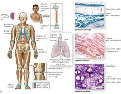
Types and Locations of Cartilage
Cartilage is a semi-rigid connective tissue found in various locations in the body. It is classified into three main types based on the composition of their extracellular matrix and fibers:
Hyaline Cartilage: Most common type; found in the nose, trachea, larynx, costal cartilages, and articular surfaces of bones.
Fibrocartilage: Contains thick collagen fibers; found in intervertebral discs, pubic symphysis, and menisci of the knee.
Elastic Cartilage: Contains elastic fibers; found in the epiglottis and external ear.
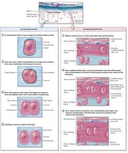
Growth of Cartilage
Cartilage grows by two mechanisms:
Interstitial Growth: Chondrocytes within the cartilage divide and secrete new matrix, expanding the cartilage from within.
Appositional Growth: New layers of cartilage are added to the surface by chondroblasts in the perichondrium.
Bone Tissue
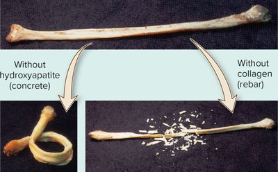
Composition of Bone
Bone is a connective tissue with a solid matrix composed of organic and inorganic components:
Organic: Collagen fibers provide flexibility and tensile strength.
Inorganic: Hydroxyapatite (calcium phosphate crystals) provides hardness and compressive strength.
The balance between these components is essential for bone function. Without collagen, bone becomes brittle; without hydroxyapatite, bone becomes flexible.
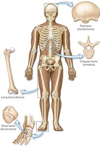
Classification of Bones
Bones are classified by shape:
Long Bones: Longer than they are wide (e.g., femur, humerus).
Short Bones: Nearly equal in length and width (e.g., tarsal bones).
Flat Bones: Thin, flat surfaces (e.g., skull, sternum).
Irregular Bones: Complex shapes (e.g., vertebrae).
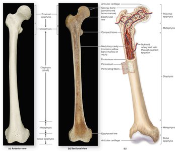
Structure of a Long Bone
Long bones have a characteristic structure:
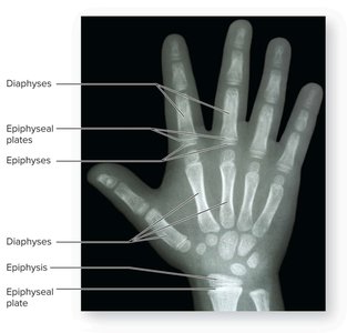
Diaphysis: Shaft of the bone.
Epiphyses: Expanded ends of the bone.
Metaphysis: Region between diaphysis and epiphysis; contains the epiphyseal plate in growing bones.
Medullary Cavity: Central cavity containing bone marrow.
Articular Cartilage: Covers joint surfaces.
Periosteum: Outer fibrous covering.
Endosteum: Lines the medullary cavity.
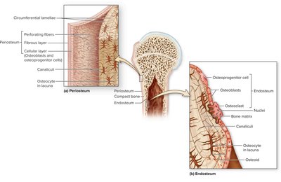
Bone Membranes
The periosteum and endosteum are connective tissue membranes that cover bone surfaces:
Periosteum: Covers external surfaces; contains osteoprogenitor cells, osteoblasts, and osteoclasts.
Endosteum: Lines internal bone surfaces; contains similar cell types as periosteum.
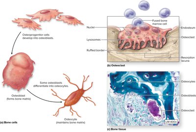
Bone Cells
Bone contains four main cell types:
Osteoprogenitor Cells: Stem cells that differentiate into osteoblasts.
Osteoblasts: Bone-forming cells; secrete bone matrix.
Osteocytes: Mature bone cells; maintain bone matrix.
Osteoclasts: Large, multinucleated cells that resorb bone.
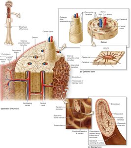
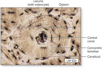
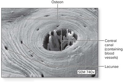
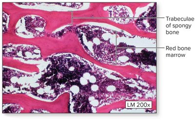
Microscopic Structure of Bone
Bone tissue is organized into two types:
Compact Bone: Dense outer layer; contains osteons (Haversian systems) with concentric lamellae around a central canal.
Spongy Bone: Internal network of trabeculae; spaces filled with bone marrow.

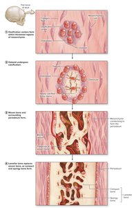
Bone Development and Growth
Ossification (Osteogenesis)
Bone formation occurs by two processes:
Intramembranous Ossification: Bone develops from a fibrous membrane; forms flat bones of the skull and clavicle.
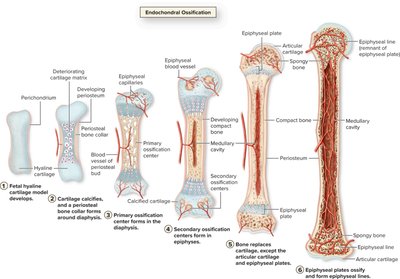
Endochondral Ossification: Bone replaces hyaline cartilage; forms most bones of the body.

Growth in Length: Epiphyseal Plate
Long bones grow in length at the epiphyseal plate, which consists of five zones:
Zone 1: Resting cartilage
Zone 2: Proliferating cartilage
Zone 3: Hypertrophic cartilage
Zone 4: Calcified cartilage
Zone 5: Ossification

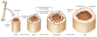
Growth in Diameter: Appositional Growth
Bones increase in diameter through appositional growth, where new bone is deposited by osteoblasts beneath the periosteum and bone is resorbed by osteoclasts on the endosteal surface.
Bone Remodeling and Repair
Bone Remodeling
Bone is continuously renewed through the coordinated actions of osteoblasts and osteoclasts. Remodeling allows bones to adapt to stress, repair microdamage, and regulate calcium levels.
Blood Supply to Bone
Bones are highly vascularized. Blood vessels enter through nutrient foramina and supply the compact and spongy bone, as well as the marrow.
Clinical Considerations
Osteoporosis
Osteoporosis is a condition characterized by decreased bone mass and increased fracture risk. It is commonly seen in older adults, especially postmenopausal women.
Summary Table: Types of Cartilage
Type | Main Fibers | Location | Function |
|---|---|---|---|
Hyaline | Collagen (Type II) | Nose, trachea, articular surfaces, costal cartilages | Support, flexibility, smooth surfaces for joints |
Fibrocartilage | Thick collagen (Type I) | Intervertebral discs, pubic symphysis, menisci | Resists compression, absorbs shock |
Elastic | Elastic fibers | Epiglottis, external ear | Flexibility, maintains shape |
