 Back
BackSkin and Body Membranes: Structure, Function, and Clinical Aspects
Study Guide - Smart Notes
 Tailored notes based on your materials, expanded with key definitions, examples, and context.
Skin and Body Membranes
Overview of Body Membranes
Body membranes are thin layers of tissue that cover surfaces, line body cavities, and form protective sheets around organs. They are classified based on their tissue composition and location in the body.
Epithelial membranes: Include cutaneous, mucous, and serous membranes.
Connective tissue membranes: Primarily represented by synovial membranes.

Epithelial Membranes
Cutaneous Membrane (Skin)
The cutaneous membrane, commonly known as the skin, is the body's largest organ and serves as a protective barrier. It consists of a superficial layer of stratified squamous epithelium (epidermis) and an underlying layer of dense fibrous connective tissue (dermis) that binds the skin to muscles or bones.
Function: Protection from mechanical damage, pathogens, and dehydration.
Structure: Epidermis (keratinized stratified squamous epithelium) and dermis (dense connective tissue).
Mucous Membranes
Mucous membranes line all body cavities that open to the exterior, such as the digestive, respiratory, urinary, and reproductive tracts. These membranes are typically moist due to the secretion of mucus, which lubricates and protects the underlying tissues.
Locations: Mouth, nasal passages, digestive tract, urinary tract, reproductive tract.
Function: Protection, secretion, and absorption.

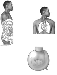
Serous Membranes
Serous membranes are composed of simple squamous epithelium and line body cavities that do not open to the exterior, such as the thoracic and abdominal cavities. They occur in pairs: the parietal layer lines the cavity wall, while the visceral layer covers the organs. A small amount of serous fluid is secreted between these layers to reduce friction.
Examples: Pleura (lungs), pericardium (heart), peritoneum (abdominal organs).
Function: Reduce friction between moving organs.
Connective Tissue Membranes
Synovial Membranes
Synovial membranes are composed of soft areolar connective tissue and lack epithelial cells. They line the fibrous capsules surrounding joints, where they secrete synovial fluid for lubrication and nutrient distribution. Synovial membranes also form bursae and tendon sheaths, which cushion and reduce friction for muscles and tendons during movement.
Locations: Joint capsules, bursae, tendon sheaths.
Function: Lubrication, shock absorption, and nutrient supply to avascular tissues.

Integumentary System
Functions of the Skin
The skin is a multifunctional organ that protects the body from external threats and helps maintain homeostasis. Its main functions include:
Protection: Against mechanical injury, pathogens, and dehydration.
Temperature regulation: Through sweating and vasodilation/vasoconstriction.
Excretion: Removal of urea and uric acid via sweat.
Sensory reception: Detection of touch, pressure, pain, and temperature.
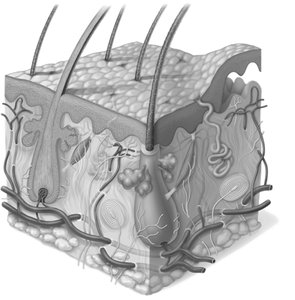
Structure of the Skin
The skin consists of three main layers: the epidermis, dermis, and hypodermis (subcutaneous layer). Each layer has distinct structural and functional properties.
Epidermis: Outermost layer, composed of keratinized stratified squamous epithelium.
Dermis: Middle layer, made of dense connective tissue, provides strength and elasticity.
Hypodermis: Deepest layer, primarily adipose tissue, provides insulation and shock absorption.

Layers of the Epidermis
The epidermis is divided into several layers, each with specialized cells and functions:
Stratum basale (basal layer): Deepest layer, actively dividing cells that replenish the upper layers.
Stratum spinosum: Thickest layer, cells contain bundles of pre-keratin filaments.
Stratum granulosum: Cells flatten, accumulate granules, and begin to lose organelles.
Stratum corneum: Outermost layer, composed of dead, keratin-filled cells that provide a tough, protective barrier.
Cutaneous Glands
Types of Skin Glands
The skin contains several types of exocrine glands, each with distinct functions:
Sudoriferous (sweat) glands: Most numerous, produce sweat for thermoregulation and waste excretion.
Sebaceous (oil) glands: Secrete sebum into hair follicles, lubricating and waterproofing the skin and hair.
Eccrine glands: Release watery sweat directly onto the skin surface.
Apocrine glands: Found in armpits and groin, secrete a thicker fluid into hair follicles, often associated with body odor.
Common Skin Disorders
Infections and Allergies
The skin is susceptible to various infections and allergic reactions, which can manifest as rashes, blisters, or lesions. Common conditions include:
Athlete's foot (tinea pedis): Fungal infection causing itchy, red, peeling skin between the toes.
Boils and carbuncles: Inflammation of hair follicles and sebaceous glands, usually due to bacterial infection.
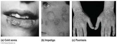
Cold sores: Fluid-filled blisters caused by herpes simplex virus, typically around the lips.
Contact dermatitis: Allergic reaction resulting in redness, swelling, and blistering after exposure to irritants like poison ivy.
Impetigo: Highly contagious bacterial infection causing pink, water-filled, crusty lesions, often around the mouth and nose.
Psoriasis: Chronic condition characterized by overproduction of skin cells, leading to red, scaly patches that may itch or bleed.
Summary Table: Types of Body Membranes
Membrane Type | Tissue Composition | Location | Main Function |
|---|---|---|---|
Cutaneous | Stratified squamous ET + dense CT | Skin (external body surface) | Protection, sensation, thermoregulation |
Mucous | Epithelium + loose CT | Lines cavities open to exterior (digestive, respiratory, urinary, reproductive tracts) | Secretion, absorption, protection |
Serous | Simple squamous ET + areolar CT | Lines closed body cavities (thoracic, abdominal) | Reduces friction, compartmentalizes organs |
Synovial | Areolar CT (no epithelium) | Lines joint cavities, bursae, tendon sheaths | Lubrication, reduces friction in joints |
