 Back
BackSkin and Body Membranes: Structure, Function, and Clinical Aspects
Study Guide - Smart Notes
 Tailored notes based on your materials, expanded with key definitions, examples, and context.
Skin and Body Membranes
Overview of Body Membranes
Body membranes are thin layers of tissue that cover surfaces, line body cavities, and form protective sheets around organs. They are classified based on their tissue composition and location in the body.
Epithelial membranes: Include cutaneous, mucous, and serous membranes.
Connective tissue membranes: Primarily represented by synovial membranes.

Epithelial Membranes
Cutaneous Membrane (Skin)
The cutaneous membrane, commonly known as the skin, is the body's largest organ and serves as a protective barrier. It consists of a superficial epidermis made of stratified squamous epithelium and an underlying layer of dense fibrous connective tissue that binds the skin to muscles or bones.
Function: Protection against mechanical damage, pathogens, and dehydration.
Structure: Epidermis (epithelial tissue) and dermis (connective tissue).
Mucous Membranes
Mucous membranes line all body cavities that open to the exterior, such as the digestive, respiratory, urinary, and reproductive tracts. These membranes are always moist due to the secretion of mucus, except in the urinary tract.
Function: Protection, secretion, and absorption.
Location: Mouth, nasal passages, digestive tract, etc.

Serous Membranes
Serous membranes are composed of simple squamous epithelium and line body cavities that do not open to the exterior, such as the thoracic and abdominal cavities. They occur in pairs:
Parietal layer: Lines the internal surface of the cavity wall.
Visceral layer: Covers the external surface of organs within the cavity.
Serous fluid: Secreted between the two layers to reduce friction.
Connective Tissue Membranes
Synovial Membranes
Synovial membranes are composed of soft areolar connective tissue and lack epithelial cells. They line the fibrous capsules surrounding joints and secrete synovial fluid for lubrication and nutrient distribution.
Function: Lubricate joints, nourish cartilage, and reduce friction.
Associated structures: Bursae sacs and tendon sheaths, which cushion and protect moving parts.

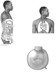
Integumentary System
Functions of the Skin
The skin is a multifunctional organ that protects the body from external threats and helps maintain homeostasis.
Protection: Against mechanical injury, pathogens, and dehydration.
Temperature regulation: Through sweating and vasodilation.
Excretion: Removal of urea and uric acid via sweat.
Sensory reception: Contains nerve endings for touch, pain, and temperature.
Structure of the Skin
The skin consists of three main layers: the epidermis, dermis, and hypodermis (subcutaneous layer).
Epidermis: Outermost layer, composed of keratinized stratified squamous epithelium.
Dermis: Dense connective tissue providing strength and elasticity; contains blood vessels, nerves, and glands.
Hypodermis: Mostly adipose tissue, providing insulation, shock absorption, and energy storage.

Layers of the Epidermis
The epidermis is divided into several distinct layers, each with specialized functions:
Stratum basale: Deepest layer; actively dividing cells that replenish the upper layers.
Stratum spinosum: Thickest layer; cells contain bundles of pre-keratin filaments.
Stratum granulosum: Cells flatten, accumulate granules, and organelles deteriorate.
Stratum corneum: Outermost layer; dead, keratin-filled cells that provide a tough, protective barrier.
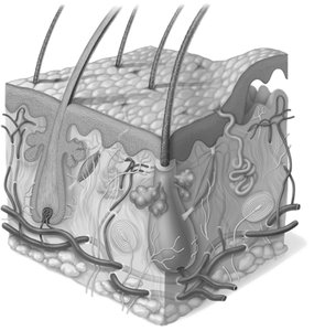
Cutaneous Glands
Types of Skin Glands
Cutaneous glands are exocrine glands located in the epidermis. They include sweat (sudoriferous) glands and sebaceous (oil) glands.
Eccrine glands: Most numerous; secrete sweat directly onto the skin surface for temperature regulation.
Apocrine glands: Found mainly in armpits and groin; secrete a thicker fluid into hair follicles, which can produce body odor.
Sebaceous glands: Secrete sebum (oil) into hair follicles to lubricate and waterproof the skin and hair.
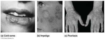
Common Skin Disorders
Infections and Allergies
The skin can be affected by various infections and allergic reactions, each with distinct clinical features:
Condition | Description | Cause |
|---|---|---|
Athlete's foot (Tinea pedis) | Itchy, red, peeling skin between toes | Fungal infection |
Boils and carbuncles | Inflammation of hair follicles and sebaceous glands | Bacterial (Staphylococcus) infection |
Cold sores | Small, fluid-filled blisters, usually around lips | Herpes simplex virus |
Contact dermatitis | Itching, redness, swelling, and blistering | Allergic reaction (e.g., poison ivy) |
Impetigo | Pink, water-filled, raised lesions with yellow crust | Highly contagious bacterial infection |
Psoriasis | Red, scaly lesions that itch, burn, and may bleed | Chronic autoimmune condition |
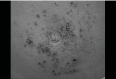
Example: Contact Dermatitis
Contact dermatitis is a common allergic reaction resulting from exposure to chemicals such as poison ivy. It is characterized by itching, redness, swelling, and sometimes blistering of the skin.
Additional info: The integumentary system also plays a role in vitamin D synthesis, immune defense, and sensory perception. Disorders of the skin can have systemic implications and may require medical intervention depending on severity.
