 Back
BackSkin and Body Membranes: Structure, Function, and Clinical Relevance
Study Guide - Smart Notes
 Tailored notes based on your materials, expanded with key definitions, examples, and context.
Skin and Body Membranes
Overview of Body Membranes
Body membranes are essential structures that cover surfaces, line body cavities, and form protective sheets around organs. They are classified based on the tissue types that compose them and serve both protective and lubricating functions.
Functions: Cover body surfaces, line cavities, and protect/lubricate organs.
Classification: Based on tissue type: epithelial membranes and connective tissue membranes.
Categories of Body Membranes
Epithelial Membranes: Also called covering and lining membranes; include cutaneous, mucous, and serous membranes.
Connective Tissue Membranes: Represented by synovial membranes.
Epithelial Membranes
General Structure
Epithelial membranes are simple organs composed of an epithelial tissue layer and an underlying connective tissue layer. They function as protective barriers and are involved in absorption and secretion.
Cutaneous Membrane (Skin)
The cutaneous membrane, or skin, is the outermost protective boundary of the body. It consists of two main layers:
Superficial Epidermis: Keratinized stratified squamous epithelium.
Underlying Dermis: Dense irregular (fibrous) connective tissue.
Characteristics: Exposed to air, dry membrane, provides protection.
Mucous Membranes (Mucosa)
Mucous membranes line all body cavities that open to the exterior. They are adapted for absorption and secretion and are typically moist due to mucus production.
Structure: Epithelium (type varies by location) and loose connective tissue (lamina propria).
Function: Absorption, secretion, and protection (e.g., respiratory and digestive tracts).
Serous Membranes (Serosa)
Serous membranes line closed ventral body cavities and occur in pairs separated by serous fluid. Each pair consists of a visceral layer (covers organs) and a parietal layer (lines cavity walls).
Structure: Simple squamous epithelium and areolar connective tissue.
Examples: Peritoneum (abdominal organs), pleurae (lungs), pericardia (heart).
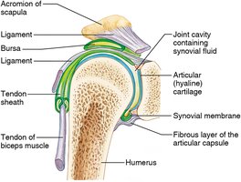
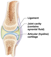
Connective Tissue Membranes
Synovial Membranes
Synovial membranes are composed solely of loose areolar connective tissue and lack epithelial components. They line the fibrous capsules surrounding joints, bursae, and tendon sheaths, secreting lubricating synovial fluid to reduce friction during movement.
Function: Cushion organs during muscle activity.
The Integumentary System
Components
The integumentary system consists of the skin (cutaneous membrane) and its appendages: sweat glands, oil glands, hair, and nails.
Functions of the Integumentary System
Protection: Acts as a barrier against mechanical, chemical, and biological hazards.
Regulation: Helps regulate body temperature and water loss.
Sensation: Contains sensory receptors for touch, pain, and temperature.
Excretion: Removes metabolic wastes through sweat.
Synthesis: Produces vitamin D in response to sunlight.
Structure of the Skin
Layers of the Skin
The skin is composed of two main layers: the epidermis and the dermis. Deep to the dermis is the subcutaneous tissue (hypodermis), which is not considered part of the skin but anchors it to underlying organs and provides insulation and shock absorption.
Epidermis: Stratified squamous epithelium; avascular; contains keratinocytes.
Dermis: Dense connective tissue; contains blood vessels, nerves, and appendages.
Hypodermis: Mostly adipose tissue; not part of the skin proper.
Layers of the Epidermis
The epidermis is composed of five layers (strata), from deepest to most superficial:
Stratum basale
Stratum spinosum
Stratum granulosum
Stratum lucidum (only in thick, hairless skin)
Stratum corneum

Specialized Cells of the Epidermis
Melanocytes: Produce melanin pigment; mostly in the stratum basale.
Epidermal dendritic cells: Activate immune responses.
Merkel cells: Associated with sensory nerve endings; function as touch receptors.
Dermis Structure
The dermis is divided into two regions:
Papillary Layer: Upper region; contains dermal papillae, capillary loops, and sensory receptors.
Reticular Layer: Deepest region; contains dense irregular connective tissue, blood vessels, glands, and pressure receptors.

Other Dermal Features
Cutaneous sensory receptors: Detect touch, pressure, temperature, and pain.
Phagocytes: Prevent microbial invasion.
Collagen and elastic fibers: Provide strength and elasticity.
Blood vessels: Aid in temperature regulation.
Skin Color
Pigments Contributing to Skin Color
Melanin: Yellow, reddish-brown, or black pigment produced by melanocytes.
Carotene: Orange-yellow pigment from certain vegetables.
Hemoglobin: Red coloring from blood cells; oxygen content affects redness.
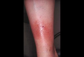
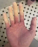
Clinical Variations in Skin Color
Redness (erythema): Due to embarrassment, inflammation, hypertension, fever, or allergy.
Pallor (blanching): Due to emotional stress, anemia, low blood pressure, or impaired blood flow.
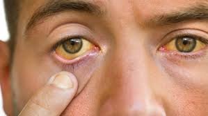
Jaundice: Yellow cast indicating liver disorder.
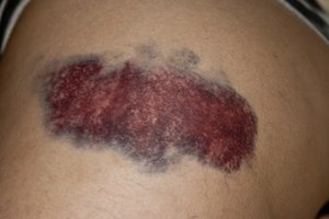
Bruising (hematomas): Black and blue marks from blood escaping into tissues.
Appendages of the Skin
Cutaneous Glands
Sebaceous (oil) glands: Produce sebum; lubricate skin and hair; kill bacteria; activated at puberty.
Sweat (sudoriferous) glands: Two types—eccrine (widely distributed, regulate temperature) and apocrine (armpits/genitals, begin at puberty, minimal role in temperature regulation).
Hair and Hair Follicles
Hair: Found almost everywhere except palms, soles, nipples, lips; produced by follicles; color from melanocytes.
Hair follicle: Inner epithelial root sheath and outer fibrous sheath; blood supply from dermal region.
Arrector pili muscle: Contracts to raise hair when cold or frightened (goosebumps).
Nails
Structure: Heavily keratinized modifications of the epidermis; colorless due to lack of pigment.
Growth: Occurs from the nail matrix beneath the nail bed.
Parts: Free edge, body, nail folds (including cuticle), root.
Body Temperature Regulation
Mechanisms of Regulation
The body maintains a narrow range of homeostatic temperature, primarily regulated by the hypothalamus. Heat is produced by oxidation of food, and over 60% escapes as heat.
Heat Loss Mechanisms: Radiation from skin, evaporation of sweat.
Heat-Promoting Mechanisms: Vasoconstriction of skin blood vessels, shivering.
Example: When body temperature rises, sweat glands increase secretion and blood vessels dilate to dissipate heat. When cold, blood vessels constrict and shivering generates heat.
