 Back
Skin and Body Membranes: Structure, Function, and Clinical Aspects
Study Guide - Smart Notes
 Tailored notes based on your materials, expanded with key definitions, examples, and context.
Skin and Body Membranes
Classification of Body Membranes
Body membranes are essential structures that cover surfaces, line cavities, and protect organs. They are classified based on tissue type into epithelial and connective tissue membranes.
Epithelial membranes: Simple organs that cover and line surfaces. Types include cutaneous, mucous, and serous membranes.
Connective tissue membranes: Include synovial membranes, which line joints and secrete lubricating fluid.
Classes of Epithelial Membranes
Cutaneous membrane: Commonly known as skin; a dry membrane forming the outermost protective boundary.
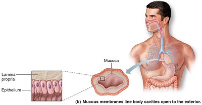
Mucous membranes: Moist membranes lining body cavities open to the exterior; adapted for absorption or secretion.
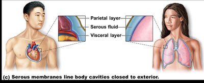
Serous membranes: Line closed body cavities; occur in pairs separated by serous fluid, with visceral and parietal layers.
Connective tissue membranes (synovial membranes) line joint capsules, bursae, and tendon sheaths, providing lubrication and cushioning.
The Integumentary System (Skin)
Functions of the Skin
The skin is a complex organ system that insulates, protects, and regulates the body. It acts as a barrier against mechanical, chemical, thermal, and microbial damage, and prevents water loss.
Insulation and cushioning: Protects deeper organs.
Protection: Guards against mechanical, chemical, thermal, UV, and microbial damage.
Regulation: Controls heat loss and retention, excretes waste, and synthesizes vitamin D.
Table: Skin Functions and How Accomplished
Functions | How accomplished |
|---|---|
Protects deeper tissues from mechanical damage (bumps) | Physical barrier contains keratin, toughens cells; fat cells cushion blows; pain receptors alert nervous system. |
Chemical damage (acids and bases) | Keratinized cells are impermeable; pain receptors alert nervous system. |
Microbe damage | Unbroken surface and acid mantle inhibit microbes; phagocytes ingest pathogens. |
Ultraviolet (UV) radiation | Melanin produced by melanocytes protects from UV damage. |
Thermal (heat or cold) damage | Contains heat/cold/pain receptors. |
Desiccation (drying out) | Water-resistant glycolipid and keratin. |
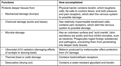
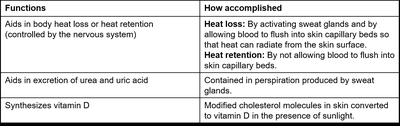
Table: Additional Skin Functions
Functions | How accomplished |
|---|---|
Aids in body heat loss or retention | Heat loss: Activates sweat glands, blood flushes into capillary beds. Heat retention: Prevents blood from flushing into capillary beds. |
Aids in excretion of urea and uric acid | Contained in perspiration produced by sweat glands. |
Synthesizes vitamin D | Modified cholesterol molecules in skin converted to vitamin D in sunlight. |
Structure of the Skin
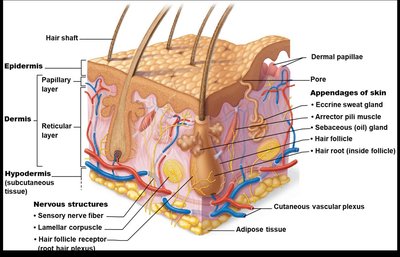
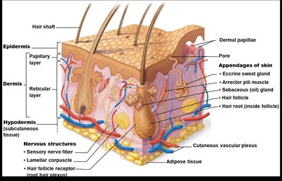
The skin is composed of two main layers: the epidermis and dermis. Beneath these lies the subcutaneous tissue (hypodermis), which anchors the skin and provides insulation.
Epidermis: Outermost layer, composed of stratified squamous epithelium; avascular.
Dermis: Dense connective tissue, contains blood vessels, nerves, and skin appendages.
Hypodermis: Mostly adipose tissue, not technically part of the integumentary system.
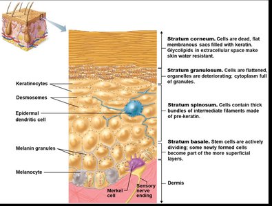
Layers of the Epidermis
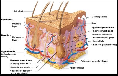
The epidermis consists of five distinct strata, each with specialized functions and cell types.
Stratum basale: Deepest layer, site of active mitosis.
Stratum spinosum: Cells become flatter and more keratinized.
Stratum granulosum: Cells are flattened, organelles deteriorate.
Stratum lucidum: Present only in thick, hairless skin (palms, soles).
Stratum corneum: Outermost layer, dead cells filled with keratin.
Cell Types in the Epidermis
Several specialized cells are found in the epidermis, each contributing to skin function and protection.
Keratinocytes: Produce keratin, a tough, protective protein.
Melanocytes: Produce melanin pigment, mostly in the stratum basale.
Epidermal dendritic cells: Activate immune responses.
Merkel cells: Associated with sensory nerve endings, serve as touch receptors.
Dermis Structure and Function
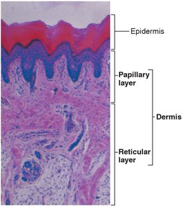
The dermis is a connective tissue layer beneath the epidermis, divided into papillary and reticular regions.
Papillary layer: Areolar connective tissue, contains dermal papillae, capillary loops, and sensory receptors.
Reticular layer: Dense irregular connective tissue, houses blood vessels, glands, and deep pressure receptors.
Skin Color
Skin color is determined by three main pigments: melanin, carotene, and hemoglobin. Variations in color can indicate physiological or pathological conditions.
Melanin: Yellow, brown, or black pigment produced by melanocytes.
Carotene: Orange-yellow pigment from diet.
Hemoglobin: Red pigment from blood cells.
Clinical color changes include erythema (redness), pallor (blanching), jaundice (yellow cast), and bruises (hematomas).
Appendages of the Skin
Skin appendages include cutaneous glands, hair, and nails, each with specialized functions.
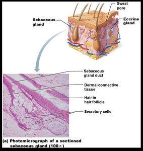
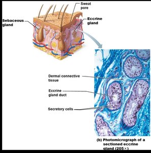
Sebaceous (oil) glands: Produce sebum, lubricate skin and hair, kill bacteria.
Sweat (sudoriferous) glands: Eccrine glands regulate temperature; apocrine glands release sweat with fatty acids and proteins.
Hair: Provides protection, produced by follicles, colored by melanin.
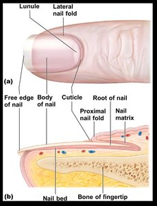
Nails: Heavily keratinized, protect fingertips, grow from nail matrix.
Homeostatic Imbalances of Skin
Infections and Allergies
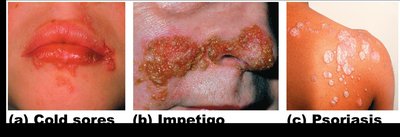
Skin is susceptible to various infections and allergic reactions, including athlete’s foot, boils, carbuncles, cold sores, contact dermatitis, impetigo, and psoriasis.
Athlete’s foot: Fungal infection causing itchy, red, peeling skin.
Boils and carbuncles: Bacterial inflammation of hair follicles.
Cold sores: Viral infection causing blisters.
Contact dermatitis: Allergic reaction to chemicals.
Impetigo: Bacterial infection with fluid-filled lesions.
Psoriasis: Chronic condition with red, scaly lesions.
Burns
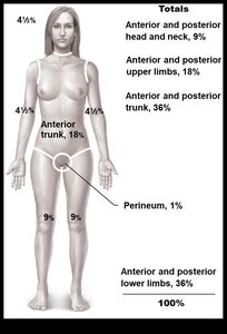
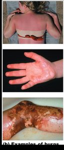
Burns cause tissue damage and are classified by depth and severity. The rule of nines estimates the extent of burns.
First-degree: Only epidermis damaged; red and swollen.
Second-degree: Epidermis and superficial dermis damaged; red, painful, blistered.
Third-degree: Destroys epidermis and dermis; area is painless, requires grafts.
Fourth-degree: Extends into deeper tissues; may require amputation.
Skin Cancer
Skin cancer is the most common form of cancer, primarily caused by UV exposure. It is classified as benign or malignant, with three main types: basal cell carcinoma, squamous cell carcinoma, and malignant melanoma.
Basal cell carcinoma: Least malignant, most common; arises from stratum basale.
Squamous cell carcinoma: Arises from stratum spinosum; can metastasize if not removed.
Malignant melanoma: Cancer of melanocytes; most deadly, rapidly metastasizes.
ABCDE Rule for Melanoma Detection
A = Asymmetry: Two sides of mole do not match.
B = Border irregularity: Borders are not smooth.
C = Color: Multiple colors present.
D = Diameter: Larger than 6 mm.
E = Evolution: Changes in any of the above characteristics.
Developmental Aspects of Skin and Body Membranes
Fetal and Aging Skin
Skin undergoes significant changes from fetal development through aging. Lanugo covers the fetus, vernix caseosa protects at birth, and milia are common in newborns. Acne appears in adolescence, and aging leads to thinning, loss of elasticity, and increased risk of skin cancer.
Lanugo: Downy hair present in fetus.
Vernix caseosa: Oily covering at birth.
Milia: Small white spots in newborns.
Aging: Skin thins, loses elasticity, balding and graying occur.
Additional info: Genetic and environmental factors contribute to skin aging and related conditions.
