 Back
BackSkin and Body Membranes: Structure, Function, and Clinical Aspects
Study Guide - Smart Notes
 Tailored notes based on your materials, expanded with key definitions, examples, and context.
Body Membranes
Classification and Function
Body membranes are thin layers of tissue that cover surfaces, line body cavities, and form protective sheets around organs. They are classified into epithelial membranes (cutaneous, mucous, serous) and connective tissue membranes (synovial).
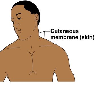
Cutaneous membrane (skin): The outermost protective boundary, exposed to air, and composed of keratinized stratified squamous epithelium overlying dense connective tissue.
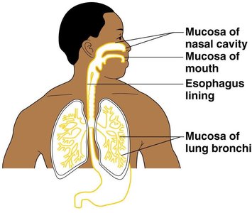
Mucous membranes: Line body cavities open to the exterior (e.g., respiratory, digestive, urinary, reproductive tracts). Adapted for absorption or secretion.
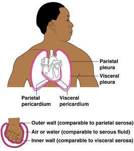
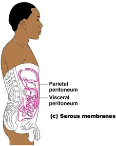
Serous membranes: Line body cavities closed to the exterior. Occur in pairs (parietal and visceral) separated by serous fluid, reducing friction between organs.
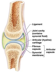
Synovial membranes: Composed only of connective tissue, line joint capsules, and secrete lubricating synovial fluid.
The Integumentary System (Skin)
Functions of the Integumentary System
The integumentary system includes the skin and its derivatives (sweat glands, oil glands, hair, nails). It serves as a barrier and performs several vital functions:
Protection: Shields underlying tissues from mechanical, chemical, and biological damage.
Regulation of body temperature: Achieved through sweat production and blood flow regulation.
Excretion: Removal of metabolic wastes via sweat.
Synthesis of vitamin D: Modified cholesterol in skin is converted to vitamin D by sunlight.
Functions | How accomplished |
|---|---|
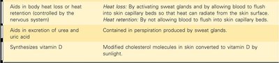
Aids in body heat loss or retention | Heat loss: By activating sweat glands and increasing blood flow to skin capillaries. Heat retention: By restricting blood flow to skin capillaries. |
Aids in excretion of urea and uric acid | Contained in perspiration produced by sweat glands. |
Synthesizes vitamin D | Modified cholesterol molecules in skin converted to vitamin D by sunlight. |

Structure of the Skin
The skin consists of three main layers: the epidermis, dermis, and subcutaneous tissue (hypodermis).
Epidermis: Outermost layer, composed of stratified squamous epithelium, often keratinized for protection.
Dermis: Dense connective tissue containing blood vessels, nerves, glands, and hair follicles.
Subcutaneous tissue (hypodermis): Not part of the skin; anchors skin to underlying organs, composed mainly of adipose tissue for shock absorption and insulation.
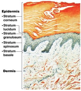
Layers of the Epidermis (Deep to Superficial: BSGLC)
Stratum basale (germinativum): Deepest layer, site of cell division.
Stratum spinosum
Stratum granulosum
Stratum lucidum: Present only in thick, hairless skin (palms, soles).
Stratum corneum: Outermost, dead, keratin-filled cells.
Layers of the Dermis
Papillary layer: Upper region with dermal papillae, capillary loops, and sensory receptors.
Reticular layer: Deepest skin layer, contains blood vessels, glands, and pressure receptors.
Collagen provides toughness; elastic fibers provide elasticity. Blood vessels help regulate temperature.
Skin Pigment and Colour Determinants
Melanin: Produced by melanocytes in the stratum basale; color ranges from yellow to black, influenced by genetics and sunlight.
Carotene: Orange-yellow pigment from vegetables.
Hemoglobin: Red pigment from blood cells in dermal capillaries; oxygen content affects redness.
Appendages of the Skin
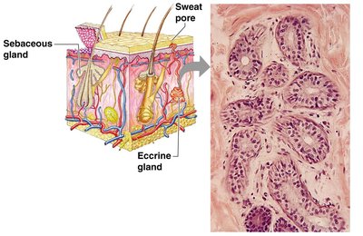
Cutaneous Glands
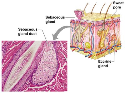
Sebaceous (oil) glands: Produce sebum, lubricates skin and hair, antibacterial, mostly empty into hair follicles, activated at puberty.
Sweat (sudoriferous) glands: Two types:
Eccrine glands: Open via duct to skin surface; produce watery sweat for temperature regulation.
Apocrine glands: Ducts empty into hair follicles; secrete sweat with fatty acids and proteins, active after puberty.
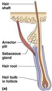
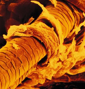
Hair, Hair Follicles, and Nails
Hair: Produced by hair follicles; consists of keratinized cells. The arrector pili muscle causes hair to stand up when cold or frightened.
Nails: Scale-like modifications of the epidermis, protect the tips of fingers and toes.
Homeostatic Imbalances of Skin
Infections and Allergies
Athlete’s foot: Fungal infection.
Boils and carbuncles: Bacterial infections.
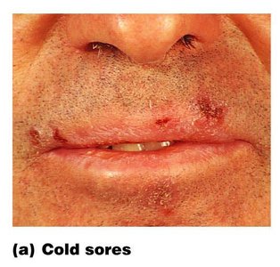
Cold sores: Viral infection (Herpes simplex virus).
Contact dermatitis: Allergic reaction.
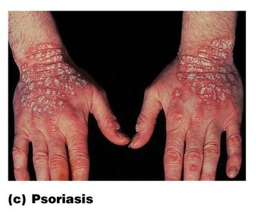
Psoriasis: Chronic condition, cause unknown, triggered by trauma, infection, or stress.
Burns
Burns are tissue damage caused by heat, electricity, UV radiation, or chemicals. Major dangers include dehydration, electrolyte imbalance, and circulatory shock.
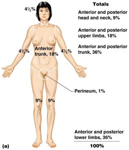
Rule of Nines: Body divided into 11 areas, each representing ~9% of total body surface area, for quick estimation of burn extent.
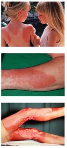
First-degree burns: Only epidermis damaged; red, swollen skin.
Second-degree burns: Epidermis and upper dermis damaged; red with blisters.
Third-degree burns: Entire skin layer destroyed; gray-white or black appearance.
Burns are considered critical if:
Over 25% of body has second-degree burns
Over 10% of body has third-degree burns
Third-degree burns involve face, hands, or feet
Skin Cancer
Skin cancer is the most common type of cancer. It is classified as benign (does not spread) or malignant (metastasizes).
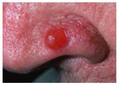
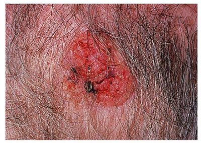
Basal cell carcinoma: Least malignant, most common, arises from stratum basale.
Squamous cell carcinoma: Can metastasize to lymph nodes, sun-induced, arises from stratum spinosum.
Malignant melanoma: Most deadly, cancer of melanocytes, rapidly metastasizes.
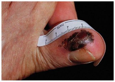
ABCD Rule for melanoma detection:
A = Asymmetry: Two sides of pigmented mole do not match.
B = Border irregularity: Borders are not smooth.
C = Color: Different colors in pigmented area.
D = Diameter: Spot is larger than 6 mm.
