 Back
BackSmooth Muscle: Structure, Function, and Comparison with Other Muscle Types
Study Guide - Smart Notes
 Tailored notes based on your materials, expanded with key definitions, examples, and context.
Smooth Muscle and Muscle Types
Histological Muscle Types: Structure and Classification
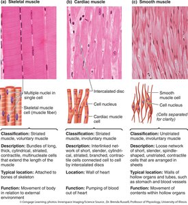
Muscle tissue in the human body is classified into three main types: skeletal, cardiac, and smooth muscle. Each type has distinct structural and functional characteristics, as well as unique modes of neurological control.
Skeletal Muscle: Striated, voluntary muscle attached to bones via tendons. Cells are large, multinucleated, and organized into fascicles. Controlled by the somatic nervous system.
Cardiac Muscle: Striated, involuntary muscle found in the heart. Cells are branched, have a single central nucleus, and are connected by intercalated discs. Controlled by the autonomic nervous system and intrinsic pacemaker activity.
Smooth Muscle: Non-striated, involuntary muscle found in walls of hollow organs and blood vessels. Cells are spindle-shaped, have a single nucleus, and are controlled by the autonomic nervous system and hormones.
Example: Skeletal muscle moves the limbs, cardiac muscle pumps blood, and smooth muscle propels food through the digestive tract.
Major Components of Smooth Muscle
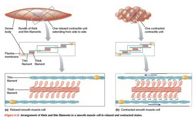
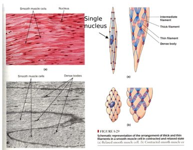
Smooth muscle cells contain specialized contractile apparatuses composed of actin (thin) and myosin (thick) filaments, as well as intermediate filaments for structural support. The arrangement of these filaments is distinct from striated muscle, allowing for unique contraction mechanics.
Thick Myosin Filaments: Myosin heads are present along the entire length, enabling extended crossbridge formation.
Thin Actin Filaments: Anchored to dense bodies, which are analogous to Z-disks in skeletal muscle.
Intermediate Filaments: Provide structural support and maintain cell shape.
Diamond-Shaped Lattice: Filaments are oriented obliquely, allowing smooth muscle to contract in multiple directions.
Example: When smooth muscle contracts, actin filaments are pulled in opposing directions, resulting in a bulging cell shape.
Smooth Muscle Contraction and Relaxation
Smooth muscle contraction is slower and more economical than skeletal muscle, allowing for sustained tension. The process is regulated primarily by calcium ions (Ca2+) and involves unique biochemical pathways.
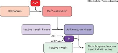
Contraction: Ca2+ enters the cell through voltage-, mechanically-, or chemically-gated channels. It binds to calmodulin, activating myosin light chain kinase (MLCK), which phosphorylates myosin, enabling crossbridge formation with actin.
Relaxation: Myosin phosphatase removes phosphate from myosin, reducing MLCK activity and leading to muscle relaxation.
Equation:
Tonic and Phasic Smooth Muscle Contractions
Types of Contraction
Smooth muscle exhibits two primary patterns of contractile activity: tonic and phasic.
Phasic Contraction: Characterized by bursts of activity and action potentials, common in hollow organs such as the digestive tract.
Tonic Contraction: Maintains continuous tension (tone) without bursts of activity, typical of blood vessel walls (tunica media).
Example: Sphincters are tonically contracted, while the intestines exhibit phasic contractions to propel contents.
Single-Unit and Multi-Unit Smooth Muscle
Functional Organization
Smooth muscle is classified based on its cellular organization and mode of excitation.
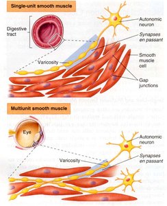
Single-Unit (Visceral) Smooth Muscle: Cells are electrically coupled via gap junctions, allowing coordinated contraction as a functional syncytium. Found in the digestive tract and uterus.
Multi-Unit Smooth Muscle: Cells operate independently and require direct stimulation by autonomic neurons. Found in the eye (ciliary muscles), large blood vessels, and airways.
Example: Single-unit smooth muscle contracts as a whole, while multi-unit smooth muscle allows finer control of contraction.
Excitation of Smooth Muscle
Mechanisms of Excitation
Smooth muscle can be excited by neural, hormonal, and physical stimuli.
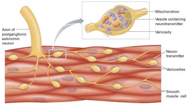
Autonomic Nervous System (ANS): Neurogenic stimulation in multi-unit muscle; modulates activity in single-unit muscle.
Hormonal: Pharmacomechanical coupling alters intracellular Ca2+ levels.
Physical: Stretch-relaxation response allows smooth muscle to develop tension when stretched, as seen in the urinary bladder.
Example: Sudden stretch causes contraction, followed by adjustment to new length (stress relaxation response).
Biochemical Response and Latch Phenomenon
Crossbridge Cycling and Energy Efficiency
Smooth muscle crossbridges are slower and more energy-efficient than those in skeletal muscle, allowing for sustained contraction (latch phenomenon). Smooth muscle does not use the creatine phosphate mechanism for energy.
Latch Phenomenon: Crossbridges remain attached for longer periods, maintaining tension with minimal ATP consumption.
Comparison: Smooth vs. Skeletal Muscle
Key Differences in Excitation and Contraction
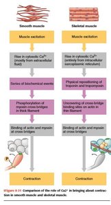
Smooth and skeletal muscle differ in their excitation mechanisms, contractile apparatus, and response to Ca2+.
Feature | Smooth Muscle | Skeletal Muscle |
|---|---|---|
Excitation | Rise in cytosolic Ca2+ (mainly extracellular) | Rise in cytosolic Ca2+ (mainly from sarcoplasmic reticulum) |
Biochemical Events | Phosphorylation of myosin crossbridges | Physical repositioning of troponin and tropomyosin |
Crossbridge Formation | Binding of actin and myosin after phosphorylation | Binding of actin and myosin after Ca2+ binding to troponin |
Contraction | Slower, sustained, economical | Rapid, short-lived, high energy consumption |
Additional info: Smooth muscle is essential for autonomic functions such as blood flow regulation, digestion, and urinary control. Its unique contractile properties allow for both rapid and sustained responses to physiological demands.
