 Back
BackSpecial Senses: Anatomy and Physiology of Vision, Hearing, Taste, and Smell
Study Guide - Smart Notes
 Tailored notes based on your materials, expanded with key definitions, examples, and context.
Special Senses: Overview
Introduction to Special Senses
The special senses include vision, hearing, equilibrium (balance), taste, and smell. These senses are mediated by specialized organs and neural pathways that allow the body to detect and interpret specific environmental stimuli.
Vision: Detected by the eyes.
Hearing and Balance: Detected by the ears.
Taste: Detected by the tongue.
Smell: Detected by the nose.
Vision
Anatomy of the Eye
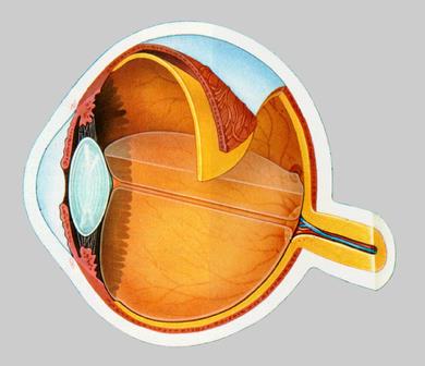
The eye is a complex organ composed of three main layers (tunics) and several specialized structures that work together to focus light and convert it into neural signals.
Fibrous Tunic: Outermost layer; includes the sclera (white of the eye) and cornea (transparent anterior part).
Vascular Tunic: Middle layer; includes the choroid, ciliary body (ciliary muscles and processes), and iris (controls pupil size).
Neural Tunic (Retina): Innermost layer; contains photoreceptors (rods and cones), the optic disk (blind spot), and the fovea centralis (area of sharpest vision).
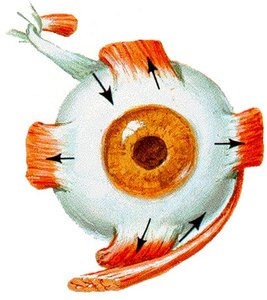
Extrinsic Eye Muscles
Six extrinsic muscles control the movement of each eye, allowing for precise tracking and positioning.
Superior rectus
Inferior rectus
Lateral rectus
Medial rectus
Superior oblique
Inferior oblique
Innervation of Eye Muscles
Oculomotor nerve (CN III): Innervates most extrinsic muscles.
Trochlear nerve (CN IV): Innervates the superior oblique muscle.
Abducens nerve (CN VI): Innervates the lateral rectus muscle.
Lens and Focusing Mechanism
The lens is a clear, flexible structure that fine-tunes the focus of light onto the retina. The shape of the lens is controlled by the ciliary muscle and suspensory ligaments.
Distance vision: Ciliary muscle relaxed, lens flattened.
Near vision: Ciliary muscle contracts, lens becomes more rounded.
Presbyopia: Age-related loss of lens flexibility, leading to difficulty focusing on close objects.
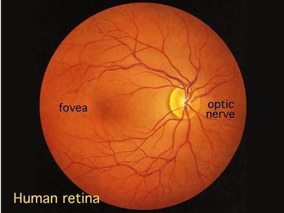
Retina and Photoreceptors
The retina contains two main types of photoreceptors:
Rods: Sensitive to low light, responsible for night vision.
Cones: Responsible for color vision and visual acuity.
Common Eye Disorders
Macular degeneration: Loss of central vision due to damage to the fovea.
Glaucoma: Increased intraocular pressure damages the optic nerve.
Retinal detachment: Separation of the retina from underlying tissue.
Hearing and Balance
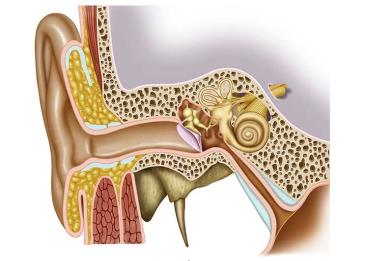
Anatomy of the Ear
The ear is divided into three main regions: external, middle, and inner ear. Each region plays a specific role in hearing and balance.
External ear: Includes the auricle and external acoustic meatus; collects sound waves.
Middle ear: Contains the tympanic membrane (eardrum) and ossicles (malleus, incus, stapes); transmits and amplifies sound.
Inner ear: Contains the cochlea (hearing) and semicircular canals (balance); filled with fluid and houses sensory receptors.
Auditory Ossicles and Muscles
Malleus (hammer), Incus (anvil), Stapes (stirrup): Transmit vibrations from the tympanic membrane to the oval window of the inner ear.
Tensor tympani (innervated by mandibular nerve) and stapedius (innervated by facial nerve): Muscles that dampen loud sounds to protect the inner ear.
Inner Ear and Vestibulocochlear Nerve
Cochlea: Spiral-shaped organ responsible for hearing.
Semicircular canals: Detect rotational movements for balance.
Vestibulocochlear nerve (CN VIII): Transmits auditory and balance information to the brain.
Taste (Gustation)
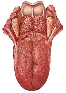
Anatomy of the Tongue
The tongue contains specialized structures called papillae, which house taste buds. Taste buds detect five primary tastes: sweet, salty, sour, bitter, and umami.
Fungiform, foliate, and circumvallate papillae: Contain taste buds.
Filiform papillae: Provide texture but do not contain taste buds.
Smell (Olfaction)
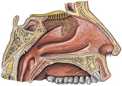
Anatomy of the Nasal Cavity
The sense of smell is mediated by olfactory receptors located in the olfactory epithelium at the roof of the nasal cavity. Olfactory nerves transmit signals to the olfactory bulb and then to the brain.
Olfactory epithelium: Contains sensory neurons for smell.
Olfactory bulb: Processes olfactory information before sending it to the brain.
Summary Table: Special Senses and Their Organs
Sense | Organ | Main Structures | Cranial Nerve |
|---|---|---|---|
Vision | Eye | Cornea, lens, retina, optic nerve | Optic (CN II) |
Hearing | Ear | Cochlea, ossicles, tympanic membrane | Vestibulocochlear (CN VIII) |
Balance | Ear | Semicircular canals, vestibule | Vestibulocochlear (CN VIII) |
Taste | Tongue | Papillae, taste buds | Facial (CN VII), Glossopharyngeal (CN IX), Vagus (CN X) |
Smell | Nose | Olfactory epithelium, olfactory bulb | Olfactory (CN I) |
Key Terms and Definitions
Photoreceptor: A specialized cell in the retina that responds to light (rods and cones).
Ossicles: The three small bones in the middle ear (malleus, incus, stapes).
Papillae: Projections on the tongue that contain taste buds.
Olfactory bulb: The brain structure that receives input from olfactory receptor neurons.
Additional info:
Special senses are distinct from general senses (such as touch and temperature) because they are localized to specific organs and have dedicated neural pathways.
Damage to the special sense organs or their neural pathways can result in significant sensory deficits.
