 Back
BackSpecial Senses: Anatomy and Physiology of the Eye and Vision
Study Guide - Smart Notes
 Tailored notes based on your materials, expanded with key definitions, examples, and context.
Special Senses
Definition and Comparison to General Senses
The special senses are distinct from general senses in both anatomical structure and function. They are responsible for complex sensory modalities such as vision, hearing, equilibrium, olfaction, and gustation.
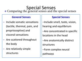
General Senses: Include somatic sensations (tactile, thermal, pain, proprioceptive) and visceral sensations; scattered throughout the body; relatively simple structures.
Special Senses: Include smell, taste, vision, hearing, and equilibrium; concentrated in specific locations in the head; anatomically distinct structures; form complex neural pathways.
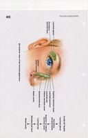
Accessory Structures of the Eye
Lacrimal Apparatus and Protective Features
The accessory structures of the eye include the eyebrows, eyelashes, eyelids, and the lacrimal apparatus, which are essential for protection, lubrication, and tear production.
Eyebrows: Located on the supraorbital ridge; help prevent sweat from entering the eye.
Eyelashes: Contain sebaceous glands at the base; protect the eye from debris; infection of these glands is called a stye.
Eyelids (palpebrae): Shade, protect, and lubricate the eye.
Lacrimal Apparatus: Consists of glands, ducts, canals, and sacs that produce and drain tears (lacrimal fluid).
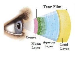
Composition of Lacrimal Fluid (Tears)
Lacrimal fluid, or tears, is a complex mixture that serves to lubricate, protect, and nourish the eye.
Oil: Provides lubrication.
Water: Maintains moisture.
Mucus: Ensures even spreading of tears.
Antibodies: Offer protection against pathogens.
Lysozyme: An antibacterial enzyme that attacks bacterial cell walls.

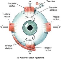
Eye Muscles
Extrinsic and Intrinsic Muscles
The eye is controlled by six extrinsic muscles that move the eyeball and one intrinsic muscle that adjusts the lens for vision.
Extrinsic Muscles: Four rectus (superior, inferior, lateral, medial) and two oblique (superior, inferior) muscles originate inside the orbit and insert on the outside surface of the eyeball.
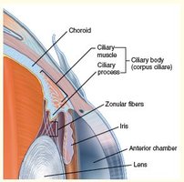
Intrinsic Muscle: The ciliary muscle originates and inserts within the eyeball, adjusting the lens for focusing.
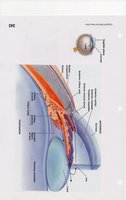
Anatomy of the Eye
Chambers and Fluids
The eye contains three main chambers: anterior, posterior, and vitreous, each filled with specific fluids that maintain eye shape and function.
Anterior Chamber: Located between the cornea and lens; contains aqueous humor.
Posterior Chamber: Located between the iris and lens.
Vitreous Chamber: Located between the lens and retina; contains vitreous humor, a jelly-like substance formed during embryonic life.
Aqueous Humor: Nourishes the lens and cornea; produced by the ciliary process and replaced every 90 minutes.
Lens: Avascular, composed of crystallins, transparent, and fine-tunes the focusing of light rays onto the retina.
Eye Disorders and Surgical Procedures
Glaucoma
Glaucoma is a condition characterized by excessive intraocular pressure, primarily due to the accumulation of aqueous humor.
Normal Pressure: 16 mmHg.
Increased Pressure: Can cause degeneration of the retina and blindness.
Diagnosis: Eye exams check for eye pressure, retina, and optic nerve health.
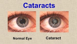
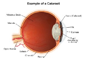
Cataracts
Cataracts involve the loss of transparency in the lens, often due to aging, injury, UV exposure, medications, or diabetes.
Symptoms: Cloudy lens, impaired vision.
Treatment: Removal of the old lens and implantation of a new artificial lens, often via phacoemulsification laser surgery.
New Lens: Contains UV protection.
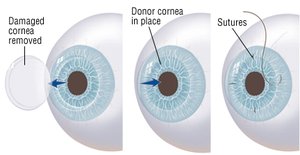
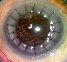
Corneal Transplants and Lasik Surgery
Corneal transplants are successful due to the avascular nature of the cornea, reducing the risk of rejection. Lasik surgery reshapes the cornea to correct refractive errors.
Corneal Transplant: Used for corneal scarring, thinning, swelling, or complications from previous surgery.
Lasik Surgery: Corrects myopia, hyperopia, and astigmatism by reshaping the cornea.
Contraindications: Not suitable for individuals under 18, pregnant, on certain medications, with unstable vision, thin cornea, poor health, dry eye syndrome, or unrealistic expectations.
Structure of the Eye: Tunics
Fibrous Tunic
The fibrous tunic consists of the cornea and sclera, providing protection and structure to the eye.
Cornea: Nonvascular, transparent coat covering the iris; allows light to enter the eye.
Sclera: The white of the eye; dense connective tissue; pierced by the optic nerve; contains the scleral venous sinus for drainage of aqueous humor.
Vascular Tunic
The vascular tunic includes the choroid, ciliary body, and iris, all of which are highly vascularized and involved in regulating light and nourishing the eye.
Choroid: Contains melanocytes and melanin, giving the eye a dark appearance.
Ciliary Body: Ciliary process secretes aqueous humor; ciliary muscle alters the shape of the lens.
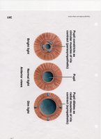
Iris: Colored portion; regulates the amount of light entering the eye; eye color is determined by chromosome 15 and melanin content.
Nervous Tunic (Retina)
The retina is the nervous tunic of the eye, responsible for detecting light and initiating neural signals for vision.
Optic Disc: The point where the optic nerve exits the eyeball.
Detached Retina: Can occur due to injury or disorders; may be treated with laser or cryosurgery.
Retinal Anatomy and Photoreceptors
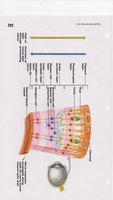
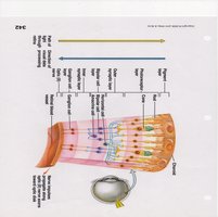
Three Layers of the Retina
The retina consists of three layers: the photoreceptor layer, bipolar layer, and ganglion cell layer, each playing a role in visual processing.
Photoreceptor Layer: Contains rods and cones that absorb light.
Bipolar Layer: Neurotransmitters released from rods and cones generate action potentials in bipolar cells.
Ganglion Cell Layer: Nerve impulses are generated and transmitted via the optic nerve.
Structure of Rods and Cones
Rods and cones are specialized photoreceptors named for their shape and function.
Rods: ~120 million; responsible for black and white vision in dim light.
Cones: ~6 million; responsible for color vision and high visual acuity in bright light.
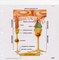
Photopigments and Color Vision
Photopigments in rods and cones absorb light and initiate the process of vision.
Rods: Contain rhodopsin (retinal + opsin).
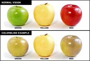
Cones: Contain idopsin; three types of opsins absorb different wavelengths (red, green, blue).
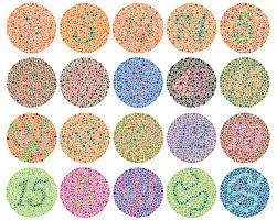
Colorblindness: Complete absence of either red or green cones; inherited as a sex-linked trait, more common in males.
Physiology of Vision
Visual Signal Processing
Vision begins with the absorption of light by photoreceptors, followed by signal transmission through bipolar and ganglion cells to the brain.
Photoreceptor Layer: Rods and cones absorb incoming light.
Bipolar Layer: Neurotransmitters released from photoreceptors generate action potentials.
Ganglion Cell Layer: Nerve impulses are generated and transmitted via the optic nerve.
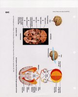
Neural Pathway: Axons of ganglion cells exit the eye, with half crossing over to the opposite side, ultimately reaching the occipital cortex (area #17) for visual processing.
Summary Table: Eye Structures and Functions
Structure | Function |
|---|---|
Cornea | Allows light entry; refracts light |
Sclera | Protects and shapes the eye |
Choroid | Nourishes retina; absorbs stray light |
Ciliary Body | Produces aqueous humor; adjusts lens shape |
Iris | Regulates light entry |
Retina | Detects light; initiates neural signals |
Lens | Focuses light onto retina |
Lacrimal Apparatus | Produces and drains tears |
Additional info: Academic context was added to clarify the physiological processes and surgical procedures, as well as to expand on the structure and function of the eye's components.
