 Back
BackSpecial Senses: Anatomy of the Visual System – Study Notes
Study Guide - Smart Notes
 Tailored notes based on your materials, expanded with key definitions, examples, and context.
Special Senses: Anatomy of the Visual System
Introduction to the Visual System
The visual system is a key component of the special senses, responsible for detecting and processing light to create visual images. It includes accessory structures, the eye itself, and neural pathways to the brain.
Accessory Eye Structures
Identification and Function
Accessory structures of the eye protect, lubricate, and support the function of the eyeball. They include:
Lacrimal apparatus: Produces and drains tears, keeping the eye moist and free of debris.
Conjunctiva: A thin, transparent membrane covering the white of the eye and lining the eyelids, providing protection and lubrication.
Palpebrae (eyelids): Protect the eye from foreign objects and help spread tears over the surface.
Medial and lateral commissures: Junctions where the eyelids meet, important for tear drainage.
Lacrimal caruncle: Contains glands producing oily secretions to lubricate the eye.
Lacrimal gland, ducts, puncta, canaliculi, sac, and nasolacrimal duct: Structures involved in tear production and drainage.
Microscopic Anatomy of the Eye
Retina and Associated Structures
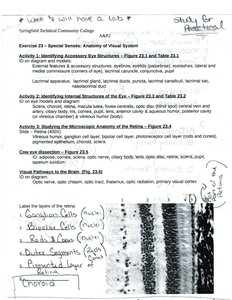
The retina is the innermost layer of the eye, responsible for detecting light and initiating neural signals. It contains several types of cells arranged in layers:
Ganglion cells: The innermost retinal neurons whose axons form the optic nerve.
Bipolar cells: Transmit signals from photoreceptors to ganglion cells.
Rods and cones: Photoreceptor cells; rods detect light intensity (black and white vision), while cones detect color.
Outer segments: Part of rods and cones containing photopigments.
Pigmented layer: Absorbs excess light and supports photoreceptors.
Choroid: Vascular layer providing oxygen and nutrients to the retina.
Gross Anatomy of the Eye
Major Structures
The eye is composed of three main layers and several important structures:
Sclera: The tough, white outer layer providing shape and protection.
Cornea: The transparent anterior part of the sclera, allowing light to enter the eye.
Choroid: Middle vascular layer supplying blood to the eye.
Retina: Inner neural layer containing photoreceptors.
Lens: Focuses light onto the retina.
Iris: Colored part of the eye, controls pupil size and light entry.
Pupil: Opening in the iris through which light passes.
Pathway of Visual Information to the Brain
Neural Pathways
Visual information is transmitted from the retina to the brain via the following pathway:
Photoreceptors (rods and cones) detect light and generate electrical signals.
Bipolar cells relay signals from photoreceptors to ganglion cells.
Ganglion cell axons converge to form the optic nerve (cranial nerve II).
Optic nerves partially cross at the optic chiasm, then continue as optic tracts to the thalamus and visual cortex.
Summary Table: Layers of the Retina
Layer | Cell Type/Function |
|---|---|
Ganglion Cell Layer | Contains ganglion cells; axons form optic nerve |
Bipolar Cell Layer | Contains bipolar neurons; relay signals from photoreceptors |
Photoreceptor Layer | Rods and cones; detect light and color |
Outer Segments | Photopigment-containing parts of rods and cones |
Pigmented Layer | Absorbs stray light, supports photoreceptors |
Choroid | Vascular layer; provides nutrients and oxygen |
Key Terms and Definitions
Photoreceptor: A specialized cell that responds to light (rods and cones).
Optic nerve: The nerve that transmits visual information from the retina to the brain.
Lacrimal apparatus: Structures involved in tear production and drainage.
Conjunctiva: Membrane covering the eye and lining the eyelids.
Additional info:
Understanding the microscopic and gross anatomy of the eye is essential for identifying normal and pathological conditions in clinical practice.
Histological identification of retinal layers is a common practical exam component in anatomy and physiology courses.
