 Back
BackSpecial Senses and Sensory Receptors: ANP Study Guide
Study Guide - Smart Notes
 Tailored notes based on your materials, expanded with key definitions, examples, and context.
Special Senses and Sensory Receptors
Overview of Sensory Perception
The somatic nervous system is responsible for processing sensory information from various organs and tissues. Sensory perception involves specialized receptors that detect stimuli and transmit signals to the brain for interpretation. This chapter focuses on the structural and functional types of sensory receptors, the anatomy of special sense organs, and the mechanisms of sensory transduction.
Types of Sensory Receptors
Sensory receptors are classified based on their structure, location, and function. These classifications help explain how different stimuli are detected and processed.
Structural Types:
Free nerve endings: Detect pain and temperature.
Encapsulated endings: Detect pressure and touch.
Specialized receptor cells: Include photoreceptors for vision.
Location Types:
Exteroceptors: Located near external stimuli (e.g., skin).
Interoceptors: Located in internal organs.
Proprioceptors: Located near moving body parts, detect position and movement.
Functional Types:
Chemoreceptors: Respond to chemical stimuli.
Osmoreceptors: Detect solute concentrations.
Nociceptors: Detect pain.
Mechanoreceptors: Respond to physical stimuli, sound, and balance.
Thermoreceptors: Detect temperature changes.
Photoreceptors: Rods and cones for vision.


Sensory Modalities
Sensory modalities refer to the different types of sensations detected by the body. These are divided into general senses and special senses.
General senses: Distributed throughout the body (e.g., touch, pain).
Special senses: Localized to specific organs (e.g., vision, hearing, taste, smell, balance).
Modality: Refers to the method by which information is encoded and perceived.
Gustation (Taste)
Gustation is the sense of taste, mediated by taste buds located within papillae on the tongue. Different tastes are transduced by specific receptor mechanisms.
Taste Types:
Sweet: Dissolved glucose, detected by G protein-coupled receptors.
Salty: Perception of sodium ions in saliva.
Sour: Detection of hydrogen ions.
Bitter: G protein-coupled receptors, often depolarize or hyperpolarize cells.
Umami: G protein-coupled receptors, perception of L-glutamate.
Taste Bud Structure:
Located within papillae (fungiform, foliate, circumvallate).
Contain gustatory receptor cells, supporting cells, and basal cells.
Innervated by facial, glossopharyngeal, and vagus nerves.

Olfaction (Smell)
Olfaction is the sense of smell, mediated by olfactory receptor neurons in the olfactory epithelium. Odorant molecules bind to receptors, initiating graded potentials and signal transduction.
Olfactory Epithelium:
Contains olfactory receptor neurons with dendrites exposed to the nasal cavity.
Odorant molecules are airborne and bind to proteins in mucus, transported to dendrites.
Neural Pathways:
Axons extend from the basal surface to the brain, connecting to the olfactory bulb.
Signals are sent to the olfactory cortex, hypothalamus, and limbic system (involved in memory and emotion).

Accessory Eye Structures
The eye is protected and supported by several accessory structures. These include glands, ducts, muscles, and protective features.
Lacrimal gland and duct: Produce and transport tears.
Extrinsic eye muscles: Four straight and two oblique muscles control eye movement.
Eyelids, eyebrows, and eyelashes: Provide protection.

Eye Anatomy
The eyeball consists of several tissue layers and specialized structures that enable vision. The main layers are the fibrous, vascular, and neural tunics.
Fibrous tunic: Sclera and cornea.
Vascular tunic: Choroid, ciliary body, iris.
Neural tunic (sensory layer): Retina, pigmented epithelium, rods and cones, optic nerve.
Lens: Biconvex, flexible structure focusing light on the retina, held by suspensory ligaments attached to ciliary muscles.


Rods and Cones
Rods and cones are the photoreceptors in the retina responsible for vision. Rods detect light intensity, while cones detect color.
Rods: Cylindrical stacks covered in proteins, more numerous, sensitive to low light, distributed away from the fovea.
Cones: Detect color (blue, green, red), concentrated in the fovea, responsible for high acuity vision.

Eye/Vision Problems
Common vision problems arise from structural differences in the eyeball or coordination between the eyes.
Hyperopia: Eyeball is short, light focuses after the retina (farsightedness).
Myopia: Eyeball is long, light focuses before the retina (nearsightedness).
Diplopia: Eyes do not focus on the same spot (double vision).
Visual Processing
Visual information is processed in the brain, with images inverted and integrated for proper positioning. The ventral stream recognizes object significance, while the dorsal stream locates objects and guides movement.
Ventral stream: Uses temporal lobe structures for object recognition.
Dorsal stream: Uses parietal lobe structures for spatial location and movement guidance.

Ear Anatomy
The ear is divided into three main regions: outer, middle, and inner ear. Each region contains structures essential for hearing and balance.
Outer ear: Auricle, external auditory meatus, external auditory canal, ceruminous glands, tympanic membrane.
Middle ear: Tympanic membrane, three ossicles (malleus, incus, stapes), oval window, air-filled cavity.
Inner ear: Vestibule, cochlea (audition), three semicircular ducts (equilibrium), perilymph and endolymph fluids.

Auditory Tube (Eustachian Tube)
The Eustachian tube connects the middle ear to the pharynx, allowing air pressure equalization and drainage during infections.
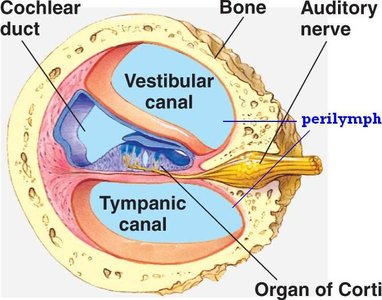
Cochlea and Hearing
The cochlea is a spiral-shaped organ responsible for auditory transduction. It contains three ducts separated by membranes and filled with perilymph or endolymph.
Scala vestibuli: Attached to oval window, filled with perilymph.
Scala media: Contains the organ of Corti, filled with endolymph, site of sensory function.
Scala tympani: Attached to round window, filled with perilymph.
Organ of Corti: Contains hair cells with stereocilia anchored to the tectorial membrane, responsible for sound transduction.


Audition (Hearing)
Hearing is a mechanical sensation. Sound waves cause the tympanic membrane to vibrate, moving ossicles and perilymph, which bends hair cells in the cochlea. This opens ion channels for depolarization, and signals are conducted through the vestibulocochlear nerve (CN VIII).
Ossicles amplify sound waves.
High frequency sounds are detected at the base of the cochlea; intensity is determined by the number of hair cells activated.
Hearing aids amplify sound; cochlear implants are used if hair cells are damaged.

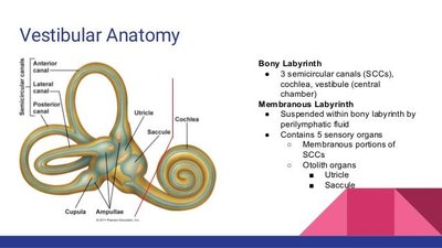
Equilibrium and Balance
Equilibrium is detected by mechanoreceptors in the vestibule and semicircular ducts. Static equilibrium is sensed in the vestibule, while dynamic equilibrium is sensed by movement in the semicircular ducts.
Cells slide and bend hairs to signal head position in space.
Perilymph fills the semicircular ducts.

Mechanoreceptor Depolarization
Both auditory and balance systems use mechanoreceptors. Hair cell movement opens channels, allowing potassium (K+) influx for depolarization. In balance, hair cells bending in one direction cause depolarization, while bending in the opposite direction causes hyperpolarization.
Summary Table: Sensory Receptor Types
Type | Stimulus | Location | Example |
|---|---|---|---|
Free nerve endings | Pain, temperature | Skin, mucous membranes | Nociceptors, thermoreceptors |
Encapsulated endings | Pressure, touch | Skin, joints | Meissner's corpuscle, Pacinian corpuscle |
Specialized receptor cell | Light, sound, chemicals | Retina, cochlea, taste buds | Photoreceptors, hair cells, gustatory cells |
Exteroceptor | External stimuli | Skin, eyes, ears | Touch, vision, hearing |
Interoceptor | Internal stimuli | Internal organs | Baroreceptors, chemoreceptors |
Proprioceptor | Body position | Muscles, tendons | Muscle spindle, Golgi tendon organ |
Key Equations
Depolarization in hair cells:
Vision optics (lens focusing):
Where is the focal length, is the object distance, and is the image distance.
Additional info: Academic context was added to clarify receptor types, neural pathways, and sensory transduction mechanisms. Images were included only when directly relevant to the explanation.
