 Back
BackSpecial Senses: Olfaction, Gustation, Hearing, and Balance
Study Guide - Smart Notes
 Tailored notes based on your materials, expanded with key definitions, examples, and context.
Special Senses
Olfaction (Smell)
The sense of smell, or olfaction, is mediated by specialized structures in the nasal cavity that detect airborne chemicals (odorants) and transduce them into neural signals interpreted by the brain as distinct odors.
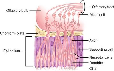
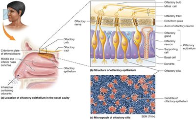
Olfactory Epithelium: Located in the roof of the nasal cavity, this epithelium contains olfactory sensory neurons, supporting cells, and basal stem cells. Olfactory neurons are bipolar, with dendrites projecting cilia into the mucus layer to increase surface area for odorant detection.
Olfactory Cilia: These structures contain receptor proteins that bind odorants, initiating the process of signal transduction.
Olfactory Nerve (Cranial Nerve I): Bundles of axons from olfactory neurons pass through the cribriform plate to synapse in the olfactory bulb, where signals are relayed to the brain.
High Turnover Rate: Olfactory neurons are replaced every 30–60 days by basal cells.
Olfactory Signal Transduction
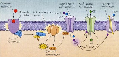
Odorant Binding: Odorant molecules must be volatile and dissolve in mucus to bind to receptor proteins on cilia.
G Protein Mechanism: Binding activates a G protein, which stimulates adenylate cyclase to produce cyclic AMP (cAMP).
Ion Channel Activation: cAMP opens cation channels, allowing Na+ and Ca2+ influx, leading to depolarization and impulse transmission.
Adaptation: Ca2+ entry contributes to adaptation, reducing sensitivity to persistent odors.
Olfactory Pathway
Olfactory receptor cells send axons through the cribriform plate to the olfactory bulb, where they synapse with mitral cells.
Mitral cell axons form the olfactory tract, projecting to the olfactory cortex and limbic system for odor perception and emotional response.
Gustation (Taste)
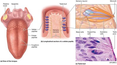
Structure of Taste Buds and Papillae
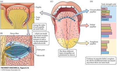
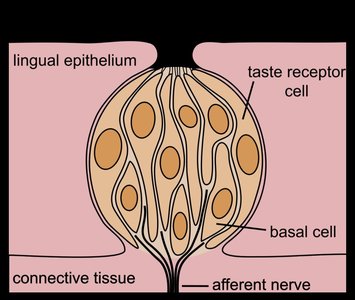
The sense of taste, or gustation, is mediated by taste buds located primarily on the tongue within papillae. Each taste bud contains receptor cells, supporting cells, and basal cells.
Papillae Types:
Fungiform: Tip of the tongue
Foliate: Sides of the tongue
Vallate (Circumvallate): Back of the tongue
Taste Buds: Contain gustatory epithelial cells (receptors), supporting cells, and basal cells (stem cells with a 7–14 day turnover).
Gustatory Hairs: Microvilli that project into the taste pore, coated in saliva, where tastants are detected.
Taste Sensations and Transduction
There are five primary taste sensations, each associated with specific chemicals and transduction mechanisms:
Sweet: Elicited by simple sugars (e.g., glucose, fructose), some artificial sweeteners, and certain toxins (e.g., ethylene glycol).
Sour: Produced by hydrogen ions (e.g., citric acid).
Salty: Elicited by metal ions (e.g., Na+, K+).
Bitter: Produced by nitrogen-containing compounds, often found in toxic substances.
Umami: Associated with amino acids, especially glutamate (e.g., in meats and broths).
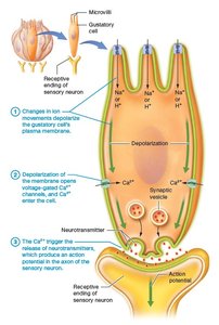
Signal Transduction in Taste
Salty: Direct influx of Na+ ions depolarizes the cell.
Sour: H+ ions open cation channels, leading to depolarization.
Sweet, Bitter, Umami: Involve G protein-coupled receptors (gustducin), which activate second messenger pathways, leading to Ca2+ release and neurotransmitter secretion.
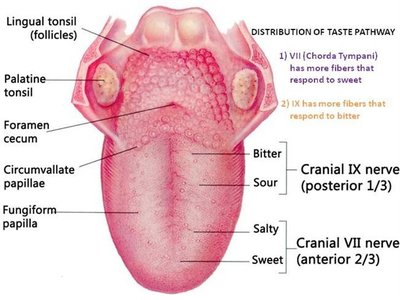
Neural Pathway for Taste
Cranial Nerves:
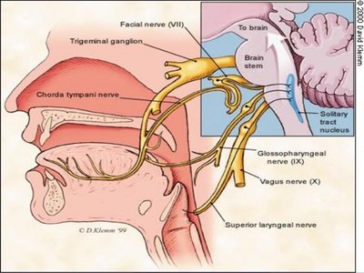
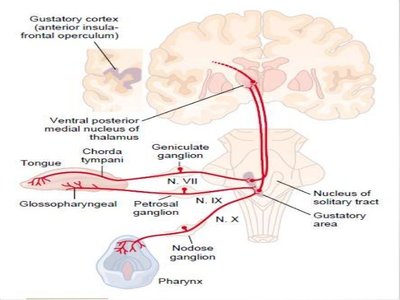
Facial Nerve (VII): Anterior two-thirds of the tongue
Glossopharyngeal Nerve (IX): Posterior one-third of the tongue
Vagus Nerve (X): Pharynx and epiglottis
Signals synapse in the solitary nucleus of the medulla, then project to the thalamus and gustatory cortex (insula).
Some fibers extend to the limbic system and hypothalamus, contributing to emotional and autonomic responses to taste.
Hearing and Balance
Anatomy of the Ear
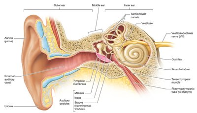
The ear is divided into three regions: external, middle, and inner ear, each with specialized structures for hearing and equilibrium.
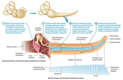
External Ear: Auricle (pinna) funnels sound into the external auditory canal, ending at the tympanic membrane (eardrum).
Middle Ear: Air-filled cavity containing the auditory ossicles (malleus, incus, stapes) that transmit and amplify sound vibrations from the tympanic membrane to the oval window.
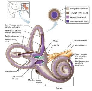
Inner Ear: Contains the bony and membranous labyrinths, filled with perilymph and endolymph, respectively. Key structures include the cochlea (hearing), vestibule, and semicircular canals (balance).
Auditory Ossicles and Sound Transmission
Ossicles: Malleus (hammer), incus (anvil), and stapes (stirrup) form a lever system that amplifies vibrations from the tympanic membrane to the oval window.
Oval Window: Transmits vibrations into the fluid-filled cochlea, where sound waves are converted into neural signals.
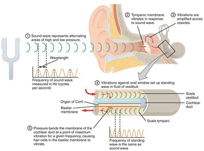
Cochlea and Sound Transduction
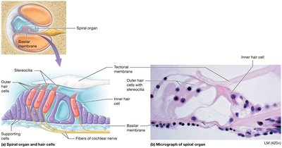
Cochlea: Spiral-shaped organ divided into three chambers: scala vestibuli (perilymph), scala media (endolymph, contains the organ of Corti), and scala tympani (perilymph).
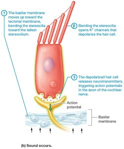
Basilar Membrane: Vibrates in response to sound waves, with different regions responding to different frequencies (base for high, apex for low).
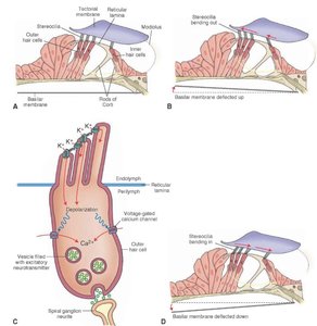
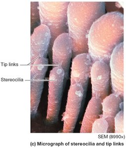
Organ of Corti: Contains inner and outer hair cells with stereocilia that transduce mechanical vibrations into electrical signals.
Properties of Sound Waves
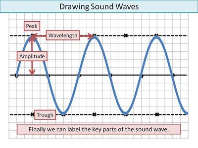
Frequency (Hz): Number of waves per second; determines pitch.
Amplitude (dB): Height of the wave; determines loudness.
Normal Hearing Range: 20–20,000 Hz; hearing loss occurs above 90 dB.
Hair Cell Transduction
Vibration of the basilar membrane bends stereocilia on hair cells against the tectorial membrane.
Bending opens K+ channels, depolarizing the cell and triggering neurotransmitter release, which generates action potentials in the cochlear nerve (part of CN VIII).
Balance (Equilibrium)
The vestibular apparatus of the inner ear detects head position, linear acceleration, and rotational movements to maintain balance.
Vestibule: Contains the utricle and saccule, each with a macula (sensory organ) that detects linear acceleration and head position via hair cells embedded in a gel matrix with otoliths (calcium carbonate crystals).
Semicircular Canals: Oriented in three planes, each contains an ampulla with a crista ampullaris (receptor for rotational acceleration).
Signal Transduction: Movement of otoliths or endolymph bends hair cell cilia, altering neurotransmitter release and modifying the firing rate of vestibular nerve fibers.
Neural Pathway: Vestibular signals travel via the vestibular nerve (division of CN VIII) to the vestibular nuclei in the brainstem or the cerebellum for integration and reflex control of eye and body movements.
Structure | Function |
|---|---|
Macula (utricle/saccule) | Detects linear acceleration and head position |
Crista ampullaris (semicircular canals) | Detects rotational acceleration |
Otoliths | Provide inertia for detecting movement |
Additional info: The integration of sensory input from the vestibular apparatus, visual system, and proprioceptors is essential for maintaining balance and posture.
