 Back
Ch 17: Special Senses: Olfaction, Gustation, Vision, Hearing, and Equilibrium
Study Guide - Smart Notes
 Tailored notes based on your materials, expanded with key definitions, examples, and context.
Special Senses
Introduction to the Special Senses
The special senses include olfaction (smell), gustation (taste), vision (sight), equilibrium (balance), and hearing (audition). These senses are mediated by specialized organs and receptor cells that detect specific environmental stimuli and transmit information to the central nervous system for processing.
Olfaction: Detection of airborne chemicals (odorants).
Gustation: Detection of dissolved chemicals (tastants) in food and liquids.
Vision: Detection of light and color for image formation.
Equilibrium: Detection of head position and movement for balance.
Hearing: Detection of sound waves (vibrations in air).
Olfaction (Smell)
Olfactory Organs and Pathways
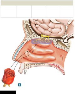
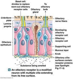
Olfactory organs are paired structures located in the nasal cavity. Each organ consists of two layers: the olfactory epithelium (containing receptor cells) and the lamina propria (containing areolar tissue, blood vessels, nerves, and olfactory glands). Olfactory glands secrete mucus that traps odorants, facilitating their interaction with receptor cells.
Olfactory Receptors: Highly modified neurons with cilia that extend into the mucus layer. These cilia contain odorant-binding proteins that interact with dissolved chemicals, generating graded and action potentials.
Olfactory Pathway: Axons of receptor neurons pass through the cribriform plate to the olfactory bulb, where they synapse with second-order neurons. These neurons project to the olfactory cortex, hypothalamus, and limbic system, bypassing the thalamus (unlike most sensory pathways).
Neurogenesis: Olfactory receptor cells are continually replaced by basal cells, making olfaction unique among special senses.
Clinical Note: The number of olfactory receptors declines with age, reducing the sense of smell.
Gustation (Taste)
Taste Buds and Papillae
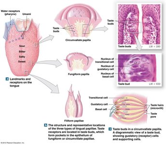
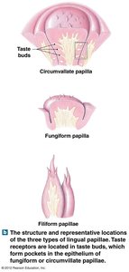
Gustation provides information about the chemical composition of food and liquids. Taste receptors (gustatory cells) are clustered in taste buds, which are located on the tongue and parts of the pharynx and larynx. Taste buds are found within specialized structures called papillae:
Circumvallate papillae: Large, located at the posterior tongue, contain many taste buds.
Fungiform papillae: Scattered across the tongue, each contains a few taste buds.
Filiform papillae: Most numerous, provide friction but do not contain taste buds.
Foliate papillae: Located on the lateral margins, contain taste buds (especially in children).
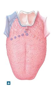
Taste Bud Structure and Neural Pathways
Gustatory Cells: Extend taste hairs through a taste pore; replaced every 10 days by basal cells.
Innervation: Taste buds are innervated by cranial nerves VII (facial, anterior 2/3 of tongue), IX (glossopharyngeal, posterior 1/3), and X (vagus, epiglottis). The trigeminal nerve (V) provides texture and temperature information.
Pathway: First-order neurons synapse in the solitary nucleus of the medulla, second-order neurons ascend to the thalamus, and third-order neurons project to the gustatory cortex in the insula.
Gustatory Discrimination
Four primary taste sensations: sweet, salty, sour, bitter.
Umami: Savory taste, sensitive to amino acids and nucleotides.
Water: Detected by receptors in the pharynx.
Vision (The Visual System)
Accessory Structures of the Eye
The eye is protected and supported by accessory structures, including the eyelids (palpebrae), conjunctiva, and the lacrimal apparatus (produces and drains tears).
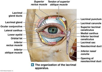
Structure of the Eyeball
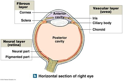
The eyeball consists of three layers and two main cavities:
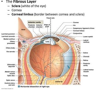
Fibrous Layer: Sclera (white of the eye) and cornea.
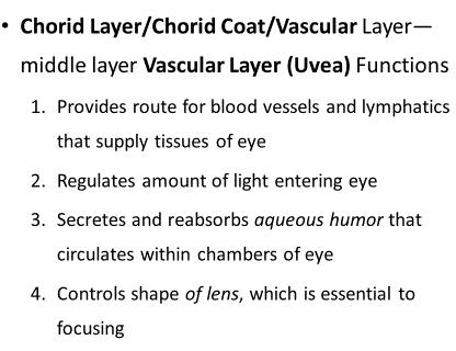
Vascular Layer (Uvea): Choroid, ciliary body, and iris. Functions include blood supply, light regulation, aqueous humor secretion, and lens shape control.
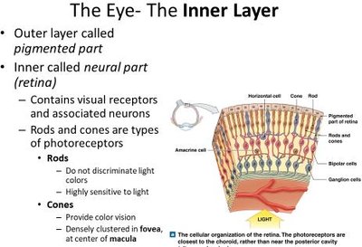
Neural Layer (Retina): Contains photoreceptors (rods and cones) and associated neurons.
Cavities: Anterior cavity (contains aqueous humor) and posterior cavity (contains vitreous humor).
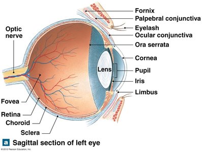
Retina and Photoreceptors
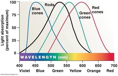
Rods: Highly sensitive to light, responsible for black-and-white vision in low light.
Cones: Responsible for color vision and visual acuity, concentrated in the fovea centralis of the macula.
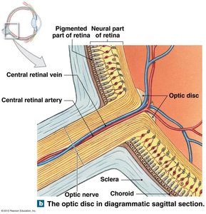
Optic Disc: Site where ganglion cell axons exit the eye as the optic nerve; no photoreceptors (blind spot).
Macula and Fovea: Region of highest visual acuity due to dense cone concentration.
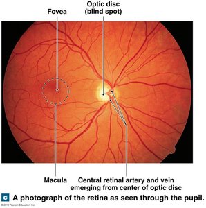
Chambers and Fluids of the Eye
Anterior Cavity: Divided into anterior and posterior chambers by the iris; contains aqueous humor, which maintains intraocular pressure and nourishes the lens and cornea.
Posterior Cavity: Contains vitreous humor, a gelatinous substance that stabilizes the eye's shape and supports the retina.
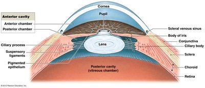
The Lens and Accommodation
Lens: Transparent, biconvex structure that focuses light onto the retina. Shape is adjusted by the ciliary body for near or distant vision (accommodation).
Accommodation: Rounder lens for near objects, flatter lens for distant objects.
Astigmatism: Irregular curvature of the cornea or lens causes distorted vision.
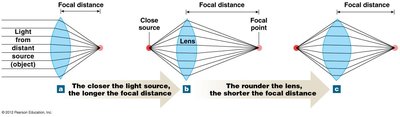
Visual Physiology
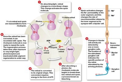
Photoreceptors: Rods and cones contain visual pigments (rhodopsin in rods; opsins in cones) that absorb light and initiate phototransduction.
Phototransduction: Light changes the shape of retinal (from 11-cis to 11-trans), activating opsin and leading to changes in membrane potential and neurotransmitter release.
Color Vision: Integration of signals from red, green, and blue cones. Color blindness results from the absence of one or more cone types.
Night Blindness: Often due to vitamin A deficiency, affecting rhodopsin regeneration.
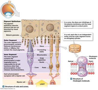
Visual Pathways
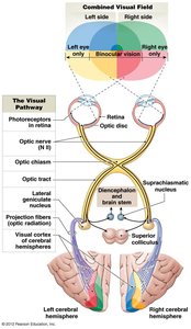
Signals from photoreceptors are relayed to bipolar cells, then to ganglion cells. Axons of ganglion cells form the optic nerve, which partially decussates at the optic chiasm.
Most fibers synapse in the lateral geniculate nucleus of the thalamus, then project to the visual cortex in the occipital lobe.
Some fibers project to other brain regions for reflexes and circadian rhythm regulation.
Hearing and Equilibrium (The Ear)
Anatomy of the Ear
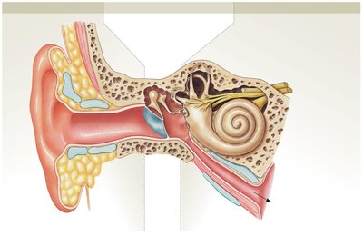
The ear is divided into three regions: external, middle, and inner ear. Each region plays a role in hearing and/or equilibrium.
External Ear: Auricle (pinna), external acoustic meatus, and tympanic membrane. Collects and directs sound waves.
Middle Ear: Tympanic cavity containing auditory ossicles (malleus, incus, stapes) that transmit vibrations to the inner ear. Connected to the nasopharynx by the auditory tube for pressure equalization.
Inner Ear: Bony and membranous labyrinths containing the cochlea (hearing), vestibule, and semicircular canals (equilibrium).
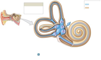
Cochlea and Auditory Transduction
Cochlea: Contains three ducts—scala vestibuli, scala tympani (both filled with perilymph), and cochlear duct (filled with endolymph). The organ of Corti (spiral organ) sits on the basilar membrane and contains hair cells (mechanoreceptors).
Sound Transmission: Sound waves vibrate the tympanic membrane, ossicles, and oval window, creating pressure waves in perilymph. These waves move the basilar membrane, stimulating hair cells against the tectorial membrane, leading to neurotransmitter release and action potentials in the cochlear nerve (part of CN VIII).
Frequency Coding: High-frequency sounds stimulate the basilar membrane near the oval window; low-frequency sounds stimulate near the apex (round window).
Intensity: Louder sounds activate more hair cells and rows.
Equilibrium: Static and Dynamic Balance
Vestibular Complex: Includes the semicircular canals (dynamic equilibrium) and the otolith organs (utricle and saccule for static equilibrium).
Semicircular Canals: Detect rotational movements. Hair cells in the ampullae are embedded in a gelatinous cupula; movement of endolymph bends the cupula, stimulating hair cells.
Otolith Organs: Detect head position and linear acceleration. Hair cells are embedded in a gelatinous matrix with calcium carbonate crystals (statoconia/otoliths) that shift with gravity, bending hair cells.
Pathways of Vestibular Sensations
Hair cells of the vestibular apparatus synapse with neurons of the vestibular branch of CN VIII. Signals are relayed to the vestibular nuclei in the brainstem, which integrate information and coordinate eye, head, and body movements for balance.
Vestibular information is also sent to the cerebellum and cerebral cortex for conscious perception of head position and movement.
Summary Table: General vs Special Senses
General Senses | Special Senses |
|---|---|
Touch, pressure, pain, temperature, proprioception | Olfaction, gustation, vision, hearing, equilibrium |
Widely distributed receptors | Localized, complex sense organs |
Simple neural pathways | Complex neural pathways, often involving cranial nerves |
