Back
BackSpecial Senses: Structure and Function of the Eye, Ear, Olfactory, and Gustatory Systems
Study Guide - Smart Notes
 Tailored notes based on your materials, expanded with key definitions, examples, and context.
Special Senses
Accessory Structures of the Eye
The accessory structures of the eye serve protective, lubricating, and functional roles to maintain vision and eye health.
Eyebrows: Shade the eye from sunlight and prevent perspiration from reaching the eyes.
Eyelids (palpebrae): Protect the eyes anteriorly.
Eyelashes: Trigger reflex blinking when touched.
Conjunctiva: Transparent mucous membrane producing lubricating mucus to prevent drying.
Lacrimal apparatus: Includes glands, canals, and ducts that produce and drain tears, supplying nutrients and removing wastes.
Extrinsic eye muscles: Control movement of the eyeball (4 rectus, 2 oblique muscles).
Conjunctiva and Its Types
The conjunctiva is a transparent mucous membrane lining the eyelids and covering the anterior surface of the eyeball, except the cornea.
Palpebral conjunctiva: Lines the eyelids.
Bulbar conjunctiva: Covers the white part of the eye, not the cornea; thin with visible blood vessels.
Extrinsic Eye Muscles and Innervation
Six extrinsic muscles control eye movement, each innervated by specific cranial nerves.
Rectus muscles: Superior (CN III), Inferior (CN III), Lateral (CN VI), Medial (CN III).
Oblique muscles: Superior (CN IV), Inferior (CN III).
Layers of the Eye Wall
The eye wall consists of three layers, each with distinct structure and function.
Fibrous layer: Outermost, dense avascular connective tissue (sclera and cornea).
Vascular layer: Middle, pigmented (choroid, ciliary body, iris).
Sensory layer: Innermost, retina (contains photoreceptors and neurons).
Fibrous Layer: Sclera and Cornea
The fibrous layer provides protection and refractive power.
Sclera: Tough, white, tendon-like; shapes the eyeball and anchors muscles.
Cornea: Transparent, allows light entry; major light-bending apparatus; stratified squamous epithelium externally, simple squamous internally; no blood vessels, rich in nerve endings.
Vascular Layer: Choroid, Ciliary Body, Iris
The vascular layer nourishes the eye and regulates light entry.
Choroid: Blood vessel-rich, dark brown; nourishes eye layers and absorbs light.
Ciliary body: Smooth muscle controls lens shape; ciliary processes secrete fluid; ciliary zonule suspends lens.
Iris: Colored part; regulates pupil size via sphincter (parasympathetic) and dilator (sympathetic) muscles.
Retina: Structure and Function
The retina is the sensory layer responsible for photoreception and signal processing.
Pigmented layer: Absorbs light, prevents scattering, acts as phagocytes, stores vitamin A.
Neural layer: Contains photoreceptors, bipolar cells, and ganglion cells; receives blood from choroid and central artery/vein.
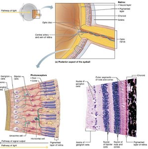
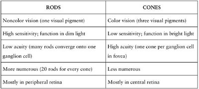
Photoreceptors: Rods and Cones
Photoreceptors transduce light energy; rods and cones have distinct functions and distributions.
Rods: Dim light and peripheral vision; high sensitivity, low acuity, noncolor vision.
Cones: Color vision, high acuity, function in bright light.
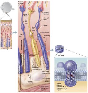
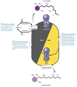
Photopigments: Retinal and Opsin
Photopigments consist of retinal (light-absorbing) and opsin (protein), with three types of opsins in cones for color vision.
Retinal: Derived from vitamin A; absorbs light.
Opsin: Glycoprotein; differs among photopigments.
Photopigment Bleaching and Regeneration
Light absorption triggers a cycle of pigment bleaching and regeneration, essential for vision.
Pigment synthesis: Rhodopsin forms in the dark.
Pigment bleaching: Light converts cis-retinal to trans-retinal, separating from opsin.
Pigment regeneration: Enzymes convert trans-retinal back to cis-retinal, rejoining opsin.
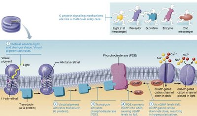
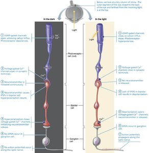
Light Transduction and Dark Current
Photoreceptor cells use cyclic GMP to maintain a dark current; light exposure hyperpolarizes the cell, inhibiting neurotransmitter release.
Dark current: Na+ channels open, depolarizing cell, continuous glutamate release.
Light exposure: Activates G-protein (transducin), closes Na+ channels, hyperpolarizes cell, reduces glutamate release.
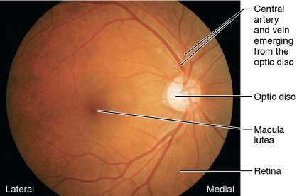
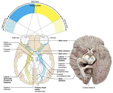
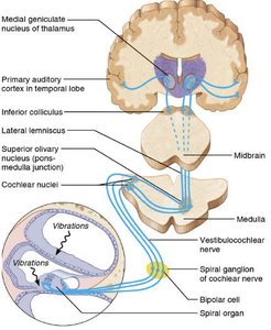
Visual Pathway: From Eye to Brain
Visual information is processed in the retina and transmitted to the brain via the optic nerve, chiasma, thalamus, and cortex.
Optic nerve: Ganglion cell axons converge at optic disc.
Optic chiasma: Medial fibers cross to opposite side.
Thalamus: Lateral geniculate nuclei relay to visual cortex.
Visual cortex: Occipital lobes process visual information.
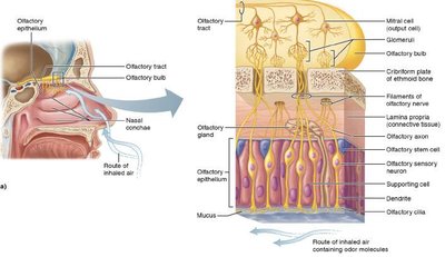
Olfactory System: Structure and Function
The olfactory epithelium contains sensory neurons, supporting cells, and stem cells for odor detection.
Olfactory sensory neurons: Bipolar neurons with cilia for odorant binding.
Supporting cells: Provide structural and metabolic support.
Olfactory stem cells: Regenerate sensory neurons.
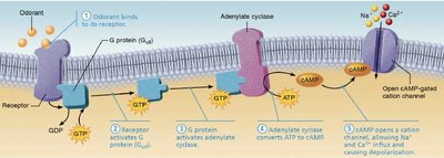
Olfactory Transduction
Odorant molecules bind to receptors, activating G-protein pathways and leading to depolarization and impulse transmission.
Odorant binds: Activates G-protein, increases cAMP.
cAMP opens cation channels: Na+ and Ca2+ influx, depolarization.
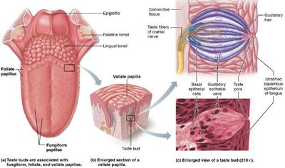
Gustatory System: Structure and Function
Taste buds are located on papillae of the tongue and contain gustatory and basal epithelial cells.
Fungiform, vallate, foliate papillae: Contain taste buds.
Gustatory epithelial cells: Receptor cells with gustatory hairs for stimulus transduction.
Basal epithelial cells: Stem cells for regeneration.
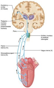
Gustatory Pathway
Taste signals are transmitted via cranial nerves VII, IX, and X to the medulla, thalamus, and gustatory cortex.
Facial nerve (VII): Anterior 2/3 of tongue.
Glossopharyngeal nerve (IX): Posterior 1/3 and pharynx.
Vagus nerve (X): Epiglottis and lower pharynx.
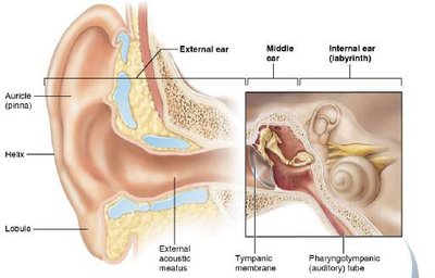
Ear Anatomy: Outer, Middle, and Inner Ear
The ear is divided into three regions, each with specialized structures for hearing and balance.
Outer ear: Auricle and external acoustic meatus.
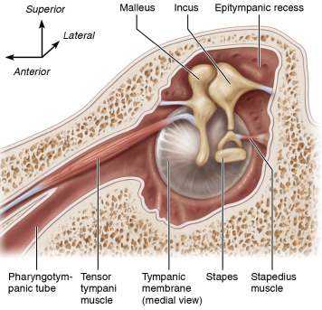
Middle ear: Tympanic membrane and auditory ossicles (malleus, incus, stapes).
Inner ear: Vestibule, cochlea, semicircular canals.
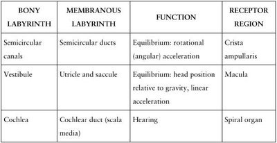
Bony and Membranous Labyrinth
The inner ear contains bony and membranous labyrinths, each with distinct functions and receptor regions.
BONY LABYRINTH | MEMBRANOUS LABYRINTH | FUNCTION | RECEPTOR REGION |
|---|---|---|---|
Semicircular canals | Semicircular ducts | Equilibrium: rotational (angular acceleration) | Crista ampullaris |
Vestibule | Utricle and saccule | Equilibrium: head position relative to gravity, linear acceleration | Macula |
Cochlea | Cochlear duct (scala media) | Hearing | Spiral organ |
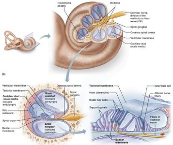
Cochlea and Spiral Organ
The cochlea is a spiral chamber divided into three chambers; the spiral organ sits on the basilar membrane and contains hair cells for sound transduction.
Scala vestibuli: Perilymph, begins at oval window.
Scala media: Endolymph, cochlear duct.
Scala tympani: Perilymph, ends at round window.
Basilar membrane: Supports spiral organ; vibrates with sound.
Tectorial membrane: Gel-like, contacts hair cell stereocilia.
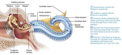
Sound Transmission and Auditory Pathway
Sound waves are transmitted from the tympanic membrane through ossicles, cochlea, and ultimately to the auditory cortex.
Tympanic membrane: Vibrates with sound waves.
Auditory ossicles: Amplify vibrations.
Pressure waves: Travel through perilymph and endolymph, vibrate basilar membrane.
Hair cells: Transduce vibrations into nerve impulses.
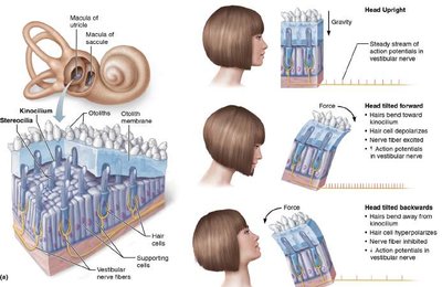
Equilibrium: Static and Dynamic
Equilibrium is maintained by the vestibular system, with maculae for static and cristae for dynamic equilibrium.
Maculae: Utricle and saccule; detect head position and linear acceleration.
Crista ampullaris: Semicircular ducts; detect rotational movement.
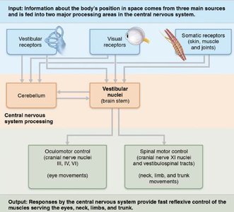
CNS Integration of Balance
Balance and orientation are integrated in the cerebellum and vestibular nuclei, using input from vestibular, somatic, and visual receptors.