 Back
BackSpecial Senses: Structure and Function of the Eye, Ear, Smell, and Taste
Study Guide - Smart Notes
 Tailored notes based on your materials, expanded with key definitions, examples, and context.
Special Senses
Overview of Special Senses
The special senses include smell, taste, sight, hearing, and equilibrium. These senses rely on specialized receptors that are often housed in large, complex sensory organs or localized clusters of cells. They provide critical information about the external environment and help maintain homeostasis.
Special sense receptors: Large, complex sensory organs or localized clusters of receptors.
Main senses: Olfaction (smell), gustation (taste), vision (sight), audition (hearing), and equilibrium (balance).
The Eye and Vision
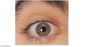
Surface Anatomy and Accessory Structures of the Eye
The eye is a highly specialized organ responsible for vision. It contains over 70% of all sensory receptors in the body, and each eye has more than one million nerve fibers transmitting information to the brain. The surface anatomy includes structures that protect and support the eye's function.
Palpebral fissure: The opening between the eyelids.
Commissures (canthi): The corners where the eyelids meet (medial and lateral).
Conjunctiva: A transparent mucous membrane covering the sclera and lining the eyelids.
Lacrimal caruncle: Contains glands that produce oily secretions.
Eyelashes and eyebrows: Protect the eye from debris and sweat.
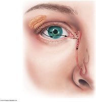
Lacrimal Apparatus
The lacrimal apparatus produces and drains tears, which lubricate and protect the eye. Tears contain mucus, antibodies, and lysozyme, an enzyme that destroys bacteria.
Lacrimal gland: Produces tears.
Lacrimal canaliculi: Drain tears from the eye surface into the lacrimal sac.
Nasolacrimal duct: Drains tears into the nasal cavity.
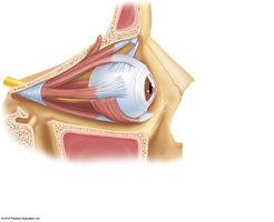
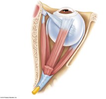
Extrinsic Muscles of the Eye
Six extrinsic muscles control the movement of each eyeball, allowing for precise and coordinated motion. These muscles originate from the bony orbit and insert onto the outer surface of the eyeball.
Superior, inferior, medial, and lateral rectus muscles: Move the eye up, down, medially, and laterally.
Superior and inferior oblique muscles: Allow for rotation and angled movements.
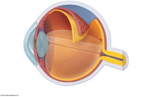
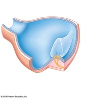
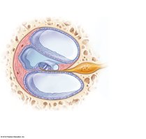
Internal Structures of the Eyeball
The eyeball is composed of three main layers (tunics) and is filled with fluids called humors. The lens divides the eye into anterior and posterior chambers.
Fibrous layer (outer): Includes the sclera (white of the eye) and cornea (transparent front part).
Vascular layer (middle): Contains the choroid, ciliary body, and iris.
Sensory layer (inner): The retina, which contains photoreceptors (rods and cones).
Humors: Aqueous humor (anterior segment) and vitreous humor (posterior segment) maintain intraocular pressure and shape.
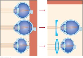
Lens: A flexible, biconvex structure that focuses light onto the retina.
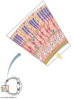
The Retina and Visual Pathways
The retina contains three major types of neurons: photoreceptors (rods and cones), bipolar cells, and ganglion cells. Light passes through these layers to stimulate the photoreceptors, which convert light into electrical signals sent to the brain via the optic nerve.
Rods: Sensitive to dim light; important for night vision.
Cones: Detect color and provide sharp vision in bright light.
Fovea centralis: Area of highest visual acuity, densely packed with cones.
Optic disc (blind spot): Where the optic nerve exits the eye; lacks photoreceptors.
Lens and Chambers of the Eye
The lens is held in place by the ciliary zonule and divides the eye into anterior and posterior segments. The anterior segment contains aqueous humor, while the posterior segment contains vitreous humor.
Lens: Focuses light onto the retina by changing shape (accommodation).
Aqueous humor: Provides nutrients to the lens and cornea; drained by the canal of Schlemm.
Vitreous humor: Maintains eye shape and holds the retina in place.

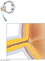
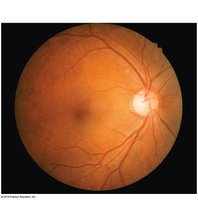
Fundus of the Eye
The fundus is the posterior wall of the eye, visible with an ophthalmoscope. It includes the macula, fovea centralis, blood vessels, and optic disc.
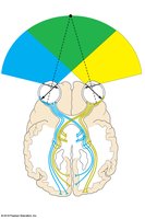
Visual Pathways to the Brain
Visual information from the retina is transmitted via the optic nerves, which partially cross at the optic chiasma, then continue as optic tracts to the thalamus and finally to the occipital lobe of the brain.
Optic nerve: Carries visual information from each eye.
Optic chiasma: Site where fibers from the nasal half of each retina cross to the opposite side.
Optic tract and radiation: Pathways to the visual cortex for image processing.
Focusing and Eye Reflexes
The eye uses several reflexes to maintain clear vision and protect itself from damage.
Convergence: Medial movement of the eyes to focus on close objects.
Photopupillary reflex: Pupil constriction in response to bright light.
Accommodation pupillary reflex: Pupil constriction when focusing on near objects.
The Ear: Hearing and Balance
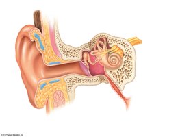
Anatomy of the Ear
The ear is divided into three regions: external, middle, and internal. It houses the organs for hearing and equilibrium, with mechanoreceptors detecting sound and head position.
External ear: Auricle (pinna) and external acoustic meatus (auditory canal).
Middle ear: Tympanic membrane (eardrum), auditory ossicles (malleus, incus, stapes), and pharyngotympanic tube.
Internal ear: Cochlea (hearing), vestibule, and semicircular canals (equilibrium).
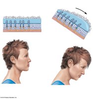
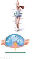
Equilibrium: Static and Dynamic
Equilibrium is maintained by receptors in the vestibule (static) and semicircular canals (dynamic) of the inner ear.
Static equilibrium: Detected by maculae in the vestibule, which sense head position relative to gravity.
Dynamic equilibrium: Detected by crista ampullaris in the semicircular canals, which sense rotational movements.
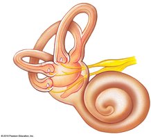
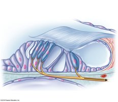
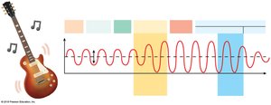
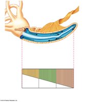
Hearing: The Cochlea and Spiral Organ of Corti
Hearing is mediated by the spiral organ of Corti, located within the cochlear duct. Hair cells on the basilar membrane are stimulated by sound vibrations, and the cochlear nerve transmits impulses to the auditory cortex.
Hair cells: Receptors for hearing, embedded in the tectorial membrane.
Basilar membrane: Supports hair cells and vibrates in response to sound.
Cochlear nerve: Sends auditory information to the brain.
Hearing and Equilibrium Deficits
Hearing loss can be classified as conduction or sensorineural deafness. Ménière’s syndrome affects both hearing and balance, causing progressive deafness and vertigo.
Conduction deafness: Impaired transmission of sound through the external or middle ear.
Sensorineural deafness: Damage to the inner ear or neural pathways.
Ménière’s syndrome: Inner ear disorder causing hearing loss and vertigo.
Chemical Senses: Smell and Taste
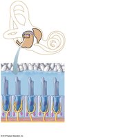
Olfaction (Smell)
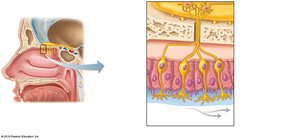
Olfactory receptors are chemoreceptors located in the nasal cavity. They are stimulated by chemicals dissolved in mucus and can detect a wide range of odors.
Olfactory epithelium: Contains receptor cells, supporting cells, and basal cells.
Olfactory nerve (cranial nerve I): Transmits signals to the olfactory bulb and brain.
Gustation (Taste)
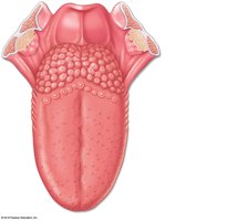
Taste buds are chemoreceptors located primarily on the tongue, but also on the soft palate and pharynx. They detect five basic tastes: sweet, sour, salty, bitter, and umami.
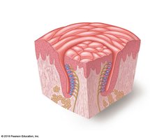
Taste buds: Found in papillae (vallate, fungiform, foliate) on the tongue.
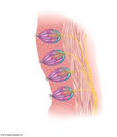
Gustatory cells: Sensory cells with microvilli (gustatory hairs) that detect dissolved chemicals.
Basal cells: Replace gustatory cells regularly.
Nerve supply: Facial (VII), glossopharyngeal (IX), and vagus (X) nerves carry taste information to the brain.
Integration of Chemical Senses
Smell and taste complement each other and often respond to the same chemical stimuli, enhancing the perception of flavors and odors.
Sense | Receptor Type | Location | Main Function |
|---|---|---|---|
Vision | Photoreceptors (rods, cones) | Retina of eye | Detect light and color |
Hearing | Hair cells (mechanoreceptors) | Cochlea of ear | Detect sound vibrations |
Equilibrium | Hair cells (mechanoreceptors) | Vestibule, semicircular canals | Detect head position and movement |
Smell | Chemoreceptors | Nasal cavity | Detect airborne chemicals |
Taste | Chemoreceptors | Taste buds on tongue | Detect dissolved chemicals |
