 Back
BackSpecial Senses: The Eye and Vision
Study Guide - Smart Notes
 Tailored notes based on your materials, expanded with key definitions, examples, and context.
Special Senses: The Eye & Vision
Overview of Special Senses
The special senses include vision, smell, hearing, taste, and equilibrium. These senses utilize specialized sensory receptors, which are distinct receptor cells localized in the head region. Vision is the most dominant sense, with approximately 70% of the body's sensory receptors located in the eye, and about half of the cerebral cortex involved in visual processing.
Anatomy of the Eye
Fibrous Layer
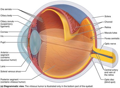
The fibrous layer is the outermost layer of the eye, composed of dense avascular connective tissue. It consists of two regions: the sclera and the cornea.
Sclera: The opaque posterior region that protects and shapes the eyeball and anchors the extrinsic eye muscles. It is continuous with the dura mater of the brain at the optic nerve exit.
Cornea: The transparent anterior one-sixth of the fibrous layer, forming a clear window that allows light to enter and bends light as it enters the eye. The cornea is covered by epithelium on both surfaces and contains numerous pain receptors, contributing to blinking and tearing reflexes.
Vascular Layer (Uvea)
The vascular layer is the middle pigmented layer of the eye, also known as the uvea. It consists of three regions:
Choroid: The posterior portion that supplies blood to all layers of the eyeball and contains brown pigment to absorb light and prevent scattering.
Ciliary Body: An anterior thickened ring of tissue surrounding the lens, containing smooth muscle bundles (ciliary muscles) that control the shape of the lens. The ciliary zonule (suspensory ligament) holds the lens in position.
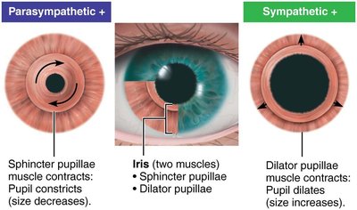
Iris: The colored part of the eye, continuous with the ciliary body, forming a circular muscular ring. The central opening, the pupil, regulates the amount of light entering the eye.
Pupil Constriction and Dilation
Pupil constriction: Sphincter pupillae (circular muscles) contract for close vision and bright light, causing pupils to constrict (parasympathetic control).
Pupil dilation: Dilator pupillae (radial muscles) contract for distant vision and dim light, causing pupils to dilate (sympathetic control).
Inner Layer (Retina)
The retina originates as an outpocketing of the brain and contains millions of photoreceptor cells, neurons, and glial cells. It is a delicate two-layered membrane:
Outer pigmented layer: Absorbs light, prevents scattering, phagocytizes photoreceptor cell fragments, and stores vitamin A.
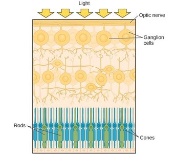
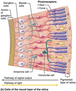
Inner neural layer: Contains photoreceptors (rods and cones), bipolar cells, and ganglion cells. Signals spread from photoreceptors to bipolar cells to ganglion cells, whose axons exit the eye as the optic nerve. The optic disc is the site where the optic nerve leaves the eye and is known as the blind spot due to the absence of photoreceptors.
Photoreceptors: Rods and Cones
The retina contains two main types of photoreceptors:
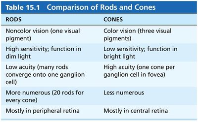
Rods: Specialized for night vision and peripheral vision. They contain a single pigment and provide vision in gray tones. Rods are more numerous and sensitive to light but do not provide color vision or sharp images. They are concentrated at the periphery of the retina.
Cones: Require bright light for activation and react more quickly than rods. They contain one of three pigments, allowing for color vision and high-resolution images. Cones are concentrated in the macula lutea, especially in the fovea centralis, which is the region of best visual acuity.
RODS | CONES |
|---|---|
Noncolor vision (one visual pigment) | Color vision (three visual pigments) |
High sensitivity; function in dim light | Low sensitivity; function in bright light |
Low acuity (many rods converge onto one ganglion cell) | High acuity (one cone per ganglion cell in fovea) |
More numerous (20 rods for every cone) | Less numerous |
Mostly in peripheral retina | Mostly in central retina |
Clinical Relevance: Retinal Detachment
Retinal detachment occurs when the pigmented and neural layers of the retina separate, allowing vitreous humor to seep between them. This can lead to permanent blindness if not treated promptly. Symptoms include a curtain-like shadow across the eye, soot-like spots, or light flashes. Treatment involves reattachment with laser surgery or vitrectomy with a gas or silicone bubble.
Lens
The lens is a biconvex, transparent, flexible, and avascular structure whose main function is to focus light on the retina. It changes shape to precisely focus light and consists of two regions: the lens epithelium (anterior cuboidal cells) and lens fibers (filled with crystallin protein). With age, the lens becomes denser, more convex, and less elastic.
Focusing Light on the Retina
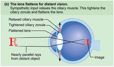
Distant vision: The eye is best adapted for distant vision. The far point of vision is the distance beyond which no change in lens shape is needed (about 20 feet for a normal eye). The ciliary muscles are relaxed, the ciliary zonule is taut, and the lens is flat.
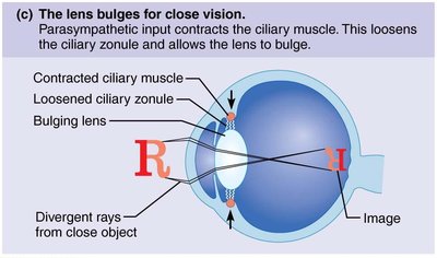
Close vision: Light from close objects diverges and requires three adjustments: accommodation of the lens (increased curvature), constriction of the pupils (to prevent divergent rays), and convergence of the eyeballs (medial rotation).
Clinical Relevance: Cataracts
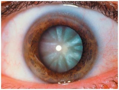
Cataracts are the clouding of the lens, often due to aging, diabetes, smoking, or exposure to intense sunlight. Crystallin proteins clump, leading to vision impairment. Cataracts can be treated by surgical replacement of the lens with an artificial one.
Light and Vision
Nature of Light
Light is a form of electromagnetic radiation, and the human eye responds only to the visible spectrum. Color perception is determined by the wavelength of light that reaches the eye.
Refraction and Lenses
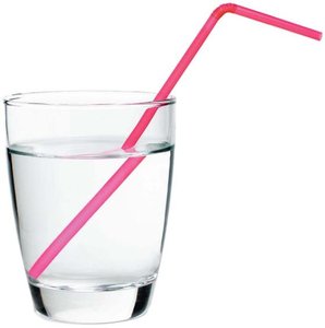
Refraction is the bending of light rays as they pass from one transparent medium to another at an oblique angle. The cornea and lens refract light to focus it on the retina. Convex lenses cause light rays to converge at a focal point, forming an image that is inverted and reversed.
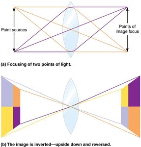
Pathway of Light Through the Eye
Light passes through the following structures in order: cornea → aqueous humor → lens → vitreous humor → neural layer of retina → photoreceptors. Light is refracted three times: entering the cornea, entering the lens, and leaving the lens. The cornea provides most of the eye's refractive power, while the lens allows for fine adjustments.
Problems of Refraction
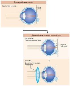
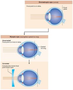
Presbyopia/Hyperopia (Farsightedness): The image focuses behind the retina due to loss of lens flexibility or a short eyeball. Distant images are clear, but close images are blurry.
Myopia (Nearsightedness): The image focuses in front of the retina due to an elongated eyeball. Nearby images are clear, but distant images are blurry.
Astigmatism: Unequal curvatures in the cornea or lens cause blurred vision.
Phototransduction and Visual Processing
Transduction of Light
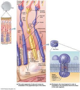
Photoreceptors (rods and cones) convert light into neural signals. Rods contain the photopigment rhodopsin, which consists of opsin (a membrane protein) and retinal (a light-absorbing molecule). When light strikes retinal, it changes shape, triggering a conformational change in opsin and initiating a signaling cascade that leads to hyperpolarization of the photoreceptor cell and transmission of visual information to the brain.
Phototransduction Process
Pigment synthesis: Rhodopsin forms and accumulates in the dark.
Pigment bleaching: Light absorption causes rhodopsin to break down into retinal and opsin.
Pigment regeneration: Rhodopsin is regenerated in the outer segments.
Cones have a similar process but require more intense light and have different types of opsins.
Light and Dark Adaptation
Light adaptation: Moving from darkness to bright light causes glare as both rods and cones are strongly stimulated. Visual acuity improves as rods turn off and cones adapt.
Dark adaptation: Moving from bright light to darkness causes temporary blindness as cones stop functioning and rods regenerate rhodopsin. Sensitivity increases over 20–30 minutes.
Visual Processing at the Retina
Visual signals travel from photoreceptors to bipolar cells to ganglion cells. Photoreceptors and bipolar cells generate graded potentials (EPSPs and IPSPs), while ganglion cells generate action potentials. In the absence of light, bipolar cells are inhibited by continuous neurotransmitter release from photoreceptors. When light hyperpolarizes photoreceptors, inhibition stops, allowing bipolar cells to depolarize and stimulate ganglion cells.
Color Vision and Visual Pathways
Opponent Process Theory
The opponent process theory explains color perception through pairs of opposing colors: red vs. green, blue vs. yellow, and black vs. white (brightness). When one color in a pair is stimulated, the other is inhibited. This theory accounts for afterimages and certain color vision phenomena.
Visual Pathway to the Brain
Axons of retinal ganglion cells form the optic nerve, which crosses at the optic chiasma and continues as optic tracts to the thalamus. Thalamic neurons project to the primary visual cortex in the occipital lobes, where conscious perception of visual images occurs.
Depth Perception
Each eye's visual field overlaps, but each eye sees objects from a slightly different angle. The visual cortex fuses these images, providing depth perception (3D vision). Loss of vision in one eye impairs depth perception, requiring reliance on other cues such as object size.
