 Back
BackSpecial Senses: Vision, Hearing, Balance, Taste, and Smell – Study Guide
Study Guide - Smart Notes
 Tailored notes based on your materials, expanded with key definitions, examples, and context.
Special Senses
Vision
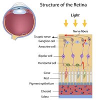
The sense of vision relies on specialized structures in the eye that detect and process light, allowing us to perceive our environment. The retina is the key sensory tissue, containing photoreceptors that convert light into neural signals.
Retina: The innermost layer of the eye, containing rods and cones, as well as several types of neurons that process visual information.
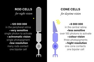
Rods vs. Cones: Rods are highly sensitive to light and enable night vision, while cones are responsible for color vision and high visual acuity.
Fovea centralis: A small region in the retina with a high density of cones, providing the sharpest vision.
Optic disc: The 'blind spot' where the optic nerve exits the eye; lacks photoreceptors.
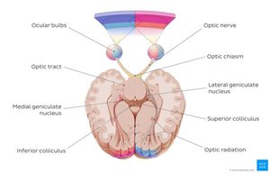
Optic nerve: Transmits visual information from the retina to the brain.
Lens, cornea, iris, pupil: Structures that focus and regulate the amount of light entering the eye.
Phototransduction
Phototransduction is the process by which light is converted into electrical signals in the retina. This process involves several steps:
Light activates rhodopsin in photoreceptors.
Retinal (a molecule within rhodopsin) changes shape.
This activates transducin (a G-protein).
Transducin activates phosphodiesterase (PDE).
PDE decreases levels of cGMP.
Low cGMP causes Na+ channels to close.
The photoreceptor hyperpolarizes.
There is a decrease in glutamate release to downstream neurons.
Key idea: Light exposure leads to less neurotransmitter release from photoreceptors.
Rods vs. Cones
Rods and cones differ in structure, function, and location:
Feature | Rods | Cones |
|---|---|---|
Light sensitivity | High | Low |
Function | Night vision | Color vision |
Location | Peripheral retina | Fovea |
Acuity | Low | High |
Retinal Cells
Photoreceptors: Detect light (rods and cones).
Bipolar cells: Transmit graded potentials from photoreceptors to ganglion cells.
Ganglion cells: Generate action potentials; their axons form the optic nerve.
Horizontal cells: Mediate lateral inhibition, enhancing contrast.
Amacrine cells: Modulate signals between bipolar and ganglion cells.
Important Concepts
Lateral inhibition: Enhances visual sharpness by inhibiting neighboring cells, increasing contrast.
Dark current: In darkness, Na+ enters photoreceptors, keeping them depolarized. In light, Na+ channels close, leading to hyperpolarization.
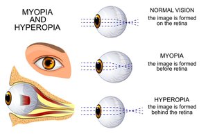
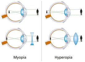
Vision Problems
Myopia (nearsightedness): Image forms in front of the retina; corrected with a concave lens.
Hyperopia (farsightedness): Image forms behind the retina; corrected with a convex lens.
Cataracts: Clouding of the lens, leading to vision loss.
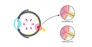
Glaucoma: Increased intraocular pressure damages the optic nerve.
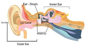
Hearing (Audition)
Hearing involves the detection of sound waves and their conversion into electrical signals by the ear. The auditory pathway transmits these signals to the brain for interpretation.
Sound Pathway
Auricle (pinna): Collects sound waves.
Tympanic membrane (eardrum): Vibrates in response to sound.
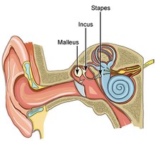
Ossicles (malleus, incus, stapes): Transmit and amplify vibrations.
Oval window: Transfers vibrations to the cochlea.
Cochlea: Contains hair cells that transduce sound into neural signals.
Hair cells → Auditory nerve (CN VIII): Send signals to the brain.
Hair Cell Physiology
Endolymph: Fluid in the cochlear duct, high in K+.
Movement of hair cells causes K+ to enter, depolarizing the cell and triggering neurotransmitter release.
Sound Encoding
Frequency (pitch): Determined by the location of stimulation on the basilar membrane (base = high frequency, apex = low frequency).
Amplitude (loudness): Determined by the size of the vibration.
Clinical Concepts
Conductive hearing loss: Due to problems in the outer or middle ear.
Sensorineural hearing loss: Due to damage to hair cells or the auditory nerve.
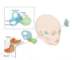
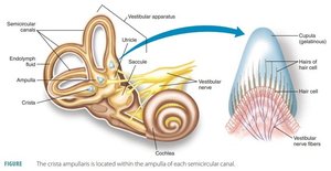
Vestibular System (Balance)
The vestibular system detects head movements and helps maintain balance and spatial orientation. It consists of structures in the inner ear that sense rotation and linear acceleration.
Semicircular canals: Detect rotational (angular) acceleration.
Utricle & saccule: Detect linear acceleration and head position relative to gravity.
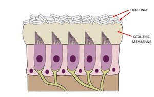
Otoliths: Calcium carbonate crystals that add mass to the otolithic membrane, aiding in the detection of movement.
Key Concept
Hair cells in the vestibular apparatus respond to fluid movement, converting mechanical stimuli into neural signals.
Clinical
Ménière’s disease: Excess endolymph causes dizziness and balance problems.
Taste (Gustation)
Taste is the detection of chemical substances (tastants) by taste buds, primarily located on the tongue. Each taste modality uses a different mechanism for signal transduction.
Taste buds: Sensory organs found in papillae on the tongue.
Taste = Chemoreception: Detection of chemicals dissolved in saliva.
Taste Types & Mechanisms
Taste | Mechanism |
|---|---|
Sweet | GPCR |
Bitter | GPCR |
Umami | GPCR |
Salty | Na+ channels |
Sour | H+ ions |
Taste adapts rapidly: Sensitivity to a taste decreases with continued exposure.
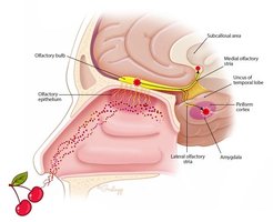
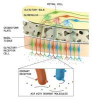
Smell (Olfaction)
Olfaction is the sense of smell, mediated by chemoreceptors in the nasal cavity. Olfactory signals are unique in that they bypass the thalamus on their way to the cortex.
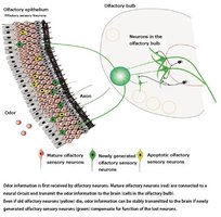
Chemoreceptors: Located in the olfactory epithelium of the nose.
Olfactory bulb: Receives input from olfactory receptor neurons and relays signals to the brain.
Direct pathway: Olfactory signals reach the cortex without first passing through the thalamus.
Study Strategy
Focus on understanding processes, not just memorizing facts.
Practice explaining concepts out loud.
Draw diagrams of key structures and pathways to reinforce learning.
