 Back
BackSpecial Senses: Vision, Hearing, Balance, Taste, and Smell – Study Guide
Study Guide - Smart Notes
 Tailored notes based on your materials, expanded with key definitions, examples, and context.
Special Senses
Overview
The special senses include vision, hearing, equilibrium (balance), taste, and smell. Each sense relies on specialized organs and receptor cells to detect environmental stimuli and transmit information to the brain for interpretation.
Vision
Key Structures of the Eye
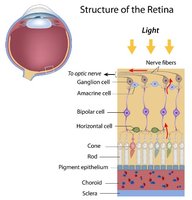
Retina: The innermost layer of the eye containing photoreceptors (rods and cones) that detect light.
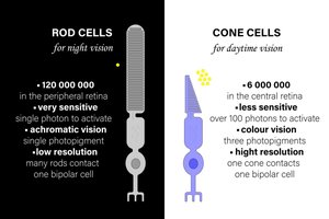
Rods: Specialized for low-light (night) vision; highly sensitive but provide low acuity and no color vision.
Cones: Responsible for color vision and high visual acuity; function best in bright light.
Fovea centralis: Area of the retina with the highest density of cones; site of sharpest vision.
Optic disc: The blind spot where the optic nerve exits the eye; lacks photoreceptors.
Optic nerve: Transmits visual information from the retina to the brain.
Lens, cornea, iris, pupil: Structures that focus and regulate the amount of light entering the eye.
Phototransduction: Steps in Light Detection
Light activates rhodopsin (a photopigment in rods).
Retinal (a molecule within rhodopsin) changes shape (isomerization).
This activates transducin (a G-protein).
Transducin activates phosphodiesterase (PDE).
PDE decreases levels of cGMP (cyclic guanosine monophosphate).
Low cGMP causes Na+ channels to close.
The photoreceptor hyperpolarizes (membrane potential becomes more negative).
Decreased glutamate release at the synapse with bipolar cells.
Key idea: In light, photoreceptors release less neurotransmitter (glutamate) than in darkness.
Rods vs. Cones
Feature | Rods | Cones |
|---|---|---|
Light Sensitivity | High | Low |
Function | Night vision | Color vision |
Location | Peripheral retina | Fovea |
Acuity | Low | High |
Retinal Cells and Signal Processing
Photoreceptors: Detect light (rods and cones).
Bipolar cells: Transmit graded potentials from photoreceptors to ganglion cells.
Ganglion cells: Generate action potentials; axons form the optic nerve.
Horizontal cells: Mediate lateral inhibition, enhancing visual contrast and sharpness.
Amacrine cells: Modulate signals between bipolar and ganglion cells.
Lateral inhibition improves visual sharpness by inhibiting neighboring cells, enhancing contrast at edges.
Dark current: In darkness, Na+ enters photoreceptors, keeping them depolarized. In light, Na+ channels close, leading to hyperpolarization.
Vision Problems
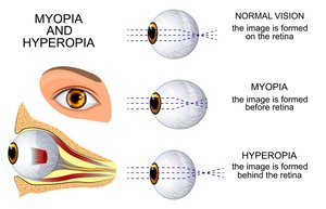
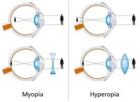
Myopia (Nearsightedness): Image focuses in front of the retina; corrected with a concave lens.
Hyperopia (Farsightedness): Image focuses behind the retina; corrected with a convex lens.
Cataracts: Clouding of the lens, leading to decreased vision.
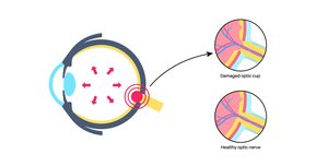
Glaucoma: Increased intraocular pressure damages the optic nerve.
Hearing (Audition)
Sound Pathway
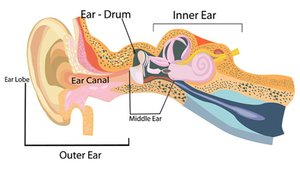
Auricle (pinna): Collects sound waves.
Tympanic membrane (eardrum): Vibrates in response to sound.
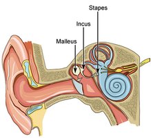
Ossicles (malleus, incus, stapes): Transmit and amplify vibrations to the oval window.
Oval window: Transfers vibrations to the cochlea.
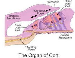
Cochlea: Contains hair cells that transduce sound into neural signals.
Hair cells: Release neurotransmitter to activate the auditory nerve (cranial nerve VIII).
Hair Cell Physiology
Endolymph: Fluid in the cochlear duct, high in K+.
Movement of the basilar membrane causes K+ to enter hair cells, depolarizing them.
Depolarization leads to neurotransmitter release and activation of the auditory nerve.
Sound Encoding
Frequency (pitch): Determined by the location of vibration on the basilar membrane (base = high frequency, apex = low frequency).
Amplitude (loudness): Determined by the size of the vibration.
Clinical Concepts
Conductive hearing loss: Caused by problems in the outer or middle ear (e.g., earwax, otitis media).
Sensorineural hearing loss: Caused by damage to hair cells or the auditory nerve.
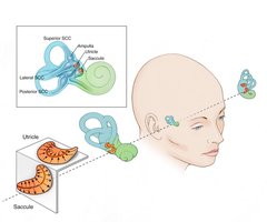
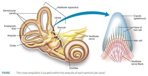
Vestibular System (Balance)
Key Structures
Semicircular canals: Detect rotational (angular) acceleration of the head.
Utricle and saccule: Detect linear acceleration and head position relative to gravity.
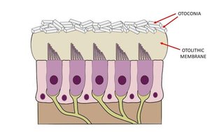
Otoliths: Calcium carbonate crystals that add mass to the otolithic membrane, aiding in the detection of movement.
Key Concept
Hair cells in the vestibular apparatus respond to movement of fluid (endolymph), which bends their stereocilia and generates nerve impulses.
Clinical
Ménière’s disease: Excess endolymph in the inner ear causes dizziness, vertigo, and balance problems.
Taste (Gustation)
Basic Facts
Taste buds are located in papillae on the tongue.
Taste is a form of chemoreception (detection of chemicals).
Taste Types & Mechanisms
Taste | Mechanism |
|---|---|
Sweet | GPCR (G-protein coupled receptor) |
Bitter | GPCR |
Umami | GPCR |
Salty | Na+ channels |
Sour | H+ ions |
Key Concept: Taste adapts rapidly, meaning sensitivity decreases with continuous exposure to a stimulus.
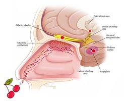
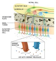
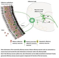
Smell (Olfaction)
Key Facts
Chemoreceptors in the nasal epithelium detect odor molecules.
Signals are transmitted to the olfactory bulb in the brain.
Olfactory signals are unique in that they initially bypass the thalamus and project directly to the cortex.
Study Strategy
Focus on understanding processes and mechanisms, not just memorization.
Practice explaining concepts out loud to reinforce learning.
Draw diagrams of key structures and pathways to visualize relationships.
