 Back
BackCh. 15 Special Senses: Vision, Olfaction, Gustation, and Hearing
Study Guide - Smart Notes
 Tailored notes based on your materials, expanded with key definitions, examples, and context.
Special Senses Overview
Introduction to Special Senses
The special senses include vision, taste, smell, hearing, and equilibrium. Unlike general senses, which are mediated by general receptors distributed throughout the body, special senses utilize distinct receptor cells localized in the head region. These specialized receptors are not simply modified nerve endings but are complex structures designed for specific sensory modalities.
Vision: Detection of light and color by the eyes.
Taste: Detection of chemical substances by taste buds.
Smell: Detection of airborne chemicals by olfactory epithelium.
Hearing: Detection of sound waves by the ear.
Equilibrium: Detection of head position and movement by the vestibular apparatus.
Vision
Accessory Structures of the Eye
The accessory structures of the eye protect and support its function. These include the eyebrows, eyelids, conjunctiva, lacrimal apparatus, and extrinsic eye muscles.
Eyebrows: Shield the eyes from sunlight and sweat.
Eyelids: Protect the eyes and help spread tears.
Conjunctiva: Transparent mucous membrane covering the front of the eye and lining the eyelids.
Lacrimal apparatus: Produces and drains tears.
Extrinsic eye muscles: Control eye movement.
of the Eyeball
The eyeball consists of three main layers and internal chambers filled with fluids. The lens divides the internal cavity into anterior and posterior segments.
Outer Fibrous Layer: Sclera and cornea.
Middle Vascular Layer: Choroid, ciliary body, iris.
Inner Nervous Layer: Retina.
Internal Cavity: Filled with humors (aqueous and vitreous).

Fibrous Layer
Sclera: Opaque, protective, shapes the eyeball, anchors muscles, continuous with dura mater.
Cornea: Transparent, allows light entry, bends light, covered by epithelium, contains sodium pumps for clarity, pain receptors for reflexes.
Vascular Layer
Choroid: Supplies blood, absorbs light to prevent scattering.
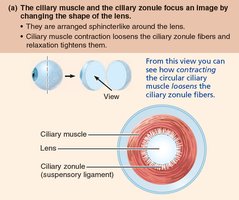
Ciliary Body: Controls lens shape, secretes fluid, holds lens in position.
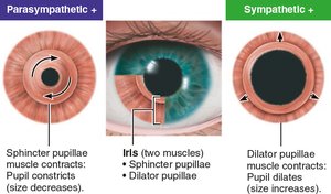
Iris: Colored part, regulates light entry via pupil.
Pupil Constriction and Dilation
Sphincter pupillae: Contracts for close vision/bright light (parasympathetic).
Dilator pupillae: Contracts for distant vision/dim light (sympathetic).
Emotional state: Pupils dilate when subject matter is appealing or requires problem-solving.
Inner Layer (Retina)
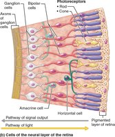
The retina is a two-layered membrane containing photoreceptors, neurons, and glial cells.
Pigmented Layer: Absorbs light, phagocytizes cell fragments, stores vitamin A.
Neural Layer: Contains photoreceptors (rods and cones), bipolar cells, ganglion cells. Signals travel from photoreceptors to bipolar cells to ganglion cells, whose axons form the optic nerve.
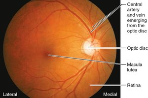
Optic Disc and Macula Lutea
Optic Disc: Site where optic nerve exits, lacks photoreceptors (blind spot).
Macula Lutea: Area with high concentration of cones, best visual acuity at fovea centralis.
Photoreceptors: Rods vs. Cones
Rods: Dim light, peripheral vision, more numerous, no color vision.
Cones: Bright light, color vision, high resolution, concentrated in macula lutea and fovea centralis.
Internal Chambers and Fluids
Posterior Segment: Contains vitreous humor, supports lens, maintains intraocular pressure.
Anterior Segment: Contains aqueous humor, supplies nutrients, drains via scleral venous sinus.
Focusing Light on the Retina
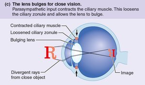
Light enters the eye through the cornea, aqueous humor, pupil, lens, and vitreous humor, reaching the retina. Refraction occurs at the cornea and lens.
Accommodation: Lens changes shape for close vision.
Presbyopia: Age-related loss of accommodation.
Visual Pathway to the Brain
Axons of retinal ganglion cells exit via the optic nerve. Medial fibers cross at the optic chiasma, forming optic tracts that carry information from the same half of the visual field. 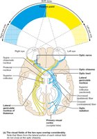
Olfaction (Smell)
Olfactory Epithelium
The olfactory epithelium is the organ of smell, located in the roof of the nasal cavity and covering the superior nasal conchae. It contains olfactory sensory neurons, supporting cells, and stem cells. 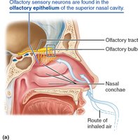
Olfactory Pathway
Olfactory nerves synapse with mitral cells in the olfactory bulbs. Mitral cells relay signals via the olfactory tract to the olfactory cortex. Some information passes through the thalamus, but olfaction is unique in that some signals go directly to the cortex. Emotional responses to odors are mediated by connections to the limbic system. 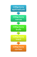
Gustation (Taste)
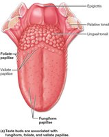
Taste Buds and Papillae
Taste buds are sensory organs for taste, mostly located on the tongue in papillae.
Fungiform papillae: Mushroom-shaped, scattered across tongue.
Foliate papillae: On side walls of tongue.
Vallate papillae: Largest, form "V" at back of tongue.
Basic Taste Sensations
There are five basic taste sensations:
Sweet: Sugars, saccharin, alcohol, some amino acids.
Sour: Hydrogen ions from acids.
Salty: Metal ions, especially sodium chloride.
Bitter: Alkaloids (quinine, nicotine, caffeine) and some nonalkaloids.
Umami: Amino acids glutamate and aspartate (meat, cheese, MSG).
Gustatory Pathway
Taste signals travel from taste buds via cranial nerves (VII, IX, X) to the solitary nucleus in the medulla, then to the thalamus and gustatory cortex. 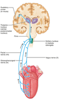
Hearing and Equilibrium
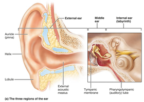
Structure of the Ear
The ear is divided into three major areas:
External (outer) ear: Auricle (pinna), external acoustic meatus, tympanic membrane.
Middle ear: Tympanic cavity, auditory ossicles (malleus, incus, stapes).
Internal (inner) ear: Bony and membranous labyrinth, vestibule, semicircular canals, cochlea.
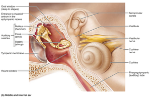
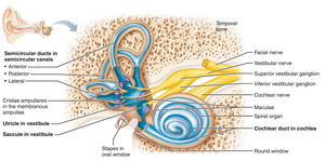
Internal Ear: Vestibule and Semicircular Canals
Vestibule: Contains saccule and utricle, houses maculae for static equilibrium.
Semicircular Canals: Three canals, each with an ampulla containing crista ampullaris for dynamic equilibrium.
Cochlea
The cochlea is a spiral chamber containing the cochlear duct and the organ of Corti, which is responsible for hearing.
Pathway of Sound Waves
Sound waves vibrate the tympanic membrane, are transmitted by ossicles to the oval window, and travel through the cochlea, stimulating hair cells. 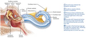
Auditory Pathway
Auditory signals travel from the cochlea via the vestibulocochlear nerve to the cochlear nuclei, then through the superior olivary nucleus, inferior colliculus, medial geniculate nucleus of the thalamus, and finally to the primary auditory cortex. 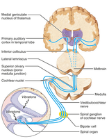
Summary Table: Comparison of Rods and Cones
Feature | Rods | Cones |
|---|---|---|
Number | More numerous | Fewer |
Location | Peripheral retina | Central retina (macula, fovea) |
Function | Dim light, peripheral vision | Bright light, color vision, high acuity |
Color Sensitivity | No | Yes |
Visual Acuity | Low | High |
Summary Table: Internal Ear Regions
Region | Main Function | Key Structures |
|---|---|---|
Vestibule | Static equilibrium | Saccule, utricle, maculae |
Semicircular Canals | Dynamic equilibrium | Ampulla, crista ampullaris |
Cochlea | Hearing | Cochlear duct, organ of Corti |
Conclusion
The special senses are essential for interacting with the environment. Each sense relies on specialized structures and pathways to detect and process specific stimuli, allowing for perception and response. Understanding these systems is fundamental for students of anatomy and physiology.
