 Back
BackSpecial Senses: Vision, Olfaction, Gustation, Hearing, and Equilibrium
Study Guide - Smart Notes
 Tailored notes based on your materials, expanded with key definitions, examples, and context.
Special Senses Overview
The special senses include vision, olfaction (smell), gustation (taste), hearing, and equilibrium. These senses are termed "special" because they have specialized receptor cells, unique neural pathways, and dedicated cortical processing areas.
Receptors: Specialized cells (not just nerve endings) act as sensory receptors.
Sensory Neurons: Bipolar neurons transmit sensory information.
Cortical Areas: Each sense has a distinct processing region in the cerebral cortex.
Vision
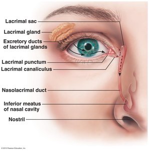
Accessory Structures of the Eye
Accessory structures protect and support the function of the eye, ensuring optimal vision and ocular health.
Eyebrows and Eyelids: Protect the eye from debris and excessive light.
Conjunctiva: Mucous membrane lining the eyelids and covering the sclera; prevents drying of the eye.
Extrinsic Eye Muscles: Six muscles control eye movement, allowing precise tracking and positioning.
Lacrimal Gland: Produces tears containing mucus, antibodies, and lysozyme for lubrication and protection.
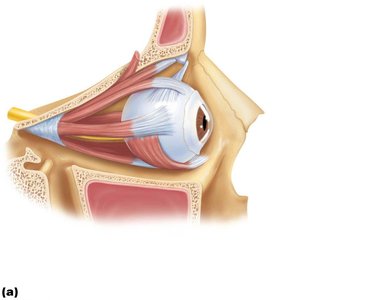
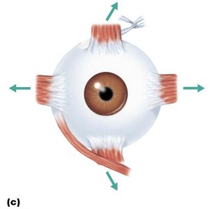
Extrinsic Eye Muscles
These muscles originate from the bony orbit and insert on the eyeball, enabling complex movements.
Four Rectus Muscles: Superior, inferior, lateral, and medial rectus move the eye in straight directions.
Two Oblique Muscles: Superior and inferior oblique muscles move the eye in the vertical plane and rotate the eyeball.
Innervation: Oculomotor (III), trochlear (IV), and abducens (VI) nerves control these muscles.
Muscle | Action | Controlling Cranial Nerve |
|---|---|---|
Lateral rectus | Moves eye laterally | VI (abducens) |
Medial rectus | Moves eye medially | III (oculomotor) |
Superior rectus | Elevates eye, turns it medially | III (oculomotor) |
Inferior rectus | Depresses eye, turns it medially | III (oculomotor) |
Inferior oblique | Elevates eye, turns it laterally | III (oculomotor) |
Superior oblique | Depresses eye, turns it laterally | IV (trochlear) |
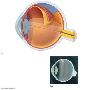
Eyeball Structure
The eyeball consists of three main tunics (layers), the lens, chambers, and the optic nerve.
Fibrous Tunic: Outermost layer; includes the sclera (white of the eye) and cornea (transparent anterior part).
Vascular Tunic: Middle layer; includes the choroid (pigmented, vascular), ciliary body (controls lens shape, secretes aqueous humor), and iris (regulates pupil size).
Sensory Tunic (Retina): Innermost layer; contains photoreceptors, bipolar cells, and ganglion cells.
Lens: Biconvex, flexible structure that focuses light on the retina.
Chambers: Anterior (aqueous humor) and posterior (vitreous humor) segments.
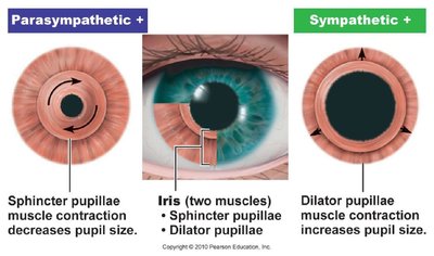
Iris and Pupil Regulation
The iris contains two muscle layers that control pupil size, regulating the amount of light entering the eye.
Sphincter Pupillae: Circular muscle; contracts to constrict the pupil (parasympathetic control).
Dilator Pupillae: Radial muscle; contracts to dilate the pupil (sympathetic control).
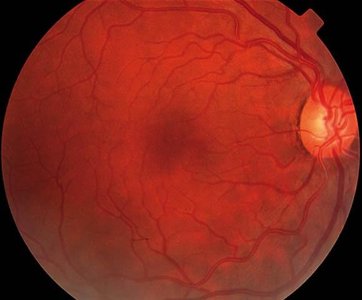
Sensory Tunic (Retina)
The retina is the site of phototransduction, converting light into neural signals.
Photoreceptors: Rods (dim light, peripheral vision) and cones (color, high acuity, central vision).
Bipolar Cells: Relay graded potentials from photoreceptors to ganglion cells.
Ganglion Cells: Generate action potentials; axons form the optic nerve.
Optic Disc: Blind spot where the optic nerve exits; lacks photoreceptors.
Fovea Centralis: Area of highest visual acuity; contains only cones.
Lens and Eye Chambers
The lens focuses light onto the retina and divides the eye into anterior and posterior chambers.
Anterior Chamber: Contains aqueous humor, nourishes lens and cornea, continually replenished.
Posterior Chamber: Contains vitreous humor, maintains eye shape, not replenished.
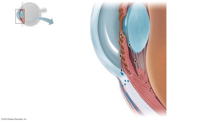
Circulation of Aqueous Humor
Aqueous humor is produced by the ciliary body, circulates through the anterior segment, and drains via the scleral venous sinus. Blockage can lead to increased intraocular pressure (glaucoma).
Visual Pathway
Light is refracted by the cornea, aqueous humor, lens, and vitreous humor before reaching the retina. Photoreceptors convert light to electrical signals, which travel via bipolar and ganglion cells to the optic nerve, cross at the optic chiasm, and reach the visual cortex in the occipital lobe.
Clinical Correlates in Vision
Conjunctivitis: Inflammation of the conjunctiva, often infectious and highly contagious.
Glaucoma: Increased intraocular pressure damages the optic nerve, leading to vision loss.
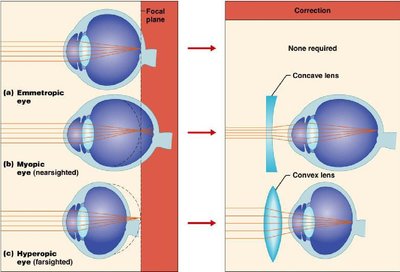
Myopia (Nearsightedness): Eyeball too long; focal point in front of retina; corrected with concave lenses.
Hyperopia (Farsightedness): Eyeball too short; focal point behind retina; corrected with convex lenses.
Astigmatism: Unequal curvature of cornea or lens; corrected with cylindrical lenses or laser surgery.
Color Blindness: Inherited lack of one or more cone pigments; more common in males (X-linked).
Olfaction (Smell)
Olfactory Epithelium and Receptors
Olfactory receptors are located in the roof of the nasal cavity and are specialized bipolar neurons. Odorants must dissolve in mucus to bind to receptors. Olfactory neurons are unique in their ability to regenerate from stem cells every 30–60 days.
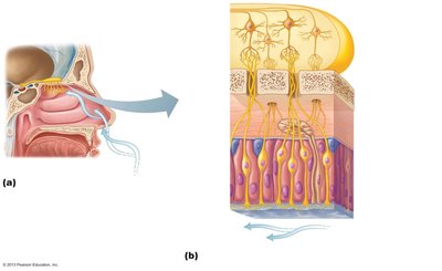
Olfactory Pathway
Olfactory receptor axons pass through the cribriform plate of the ethmoid bone, synapse in the olfactory bulb, and travel via the olfactory tract to the temporal lobe, limbic system, and hypothalamus.
Gustation (Taste)
Taste Buds and Sensations
Taste buds, primarily on the tongue, contain gustatory cells that detect tastants dissolved in saliva. The five basic tastes are sweet, sour, salty, bitter, and umami. Taste adapts quickly and is less sensitive than smell.
Threshold: Bitter has the lowest threshold (most sensitive).
Loss of Smell: Reduces taste sensation by up to 80%.
Gustatory Pathway
Taste signals travel via the facial (VII), glossopharyngeal (IX), and vagus (X) nerves to the thalamus and then to the gustatory cortex in the insula lobe.
Hearing and Equilibrium
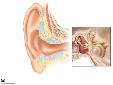
Anatomy of the Ear
The ear is divided into three regions: outer, middle, and inner ear, each with specialized structures for hearing and balance.
Outer Ear: Pinna (auricle), external auditory canal, tympanic membrane.
Middle Ear: Air-filled cavity with ossicles (malleus, incus, stapes), eustachian tube, oval and round windows.
Inner Ear: Fluid-filled; contains cochlea (hearing), vestibule, and semicircular canals (equilibrium).
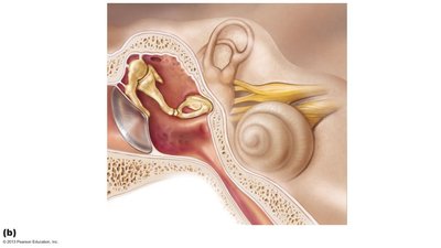
Inner Ear: Cochlea and Vestibular Apparatus
The cochlea contains the organ of Corti, the receptor for hearing. The vestibule and semicircular canals contain receptors for static and dynamic equilibrium, respectively.
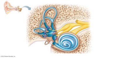
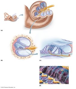
Auditory Pathway
Sound waves enter the external auditory canal, vibrate the tympanic membrane, and are transmitted via ossicles to the oval window. Fluid waves in the cochlea stimulate hair cells in the organ of Corti, generating action potentials that travel via the cochlear nerve to the auditory cortex.
Equilibrium Pathways
Vestibular signals travel via the vestibular branch of the vestibulocochlear nerve to the brainstem and cerebellum, coordinating reflexes and balance.
Clinical Correlates in Hearing
Conduction Deafness: Damage to structures conducting sound (e.g., tympanic membrane, ossicles).
Sensorineural Deafness: Damage to neural elements (hair cells, cochlea, cochlear nerve).
Additional info: The notes above expand on the provided content with definitions, clinical context, and logical organization for clarity and completeness.
