 Back
Special Senses: Vision, Smell, Taste, Hearing, and Balance – ANP College Study Notes
Study Guide - Smart Notes
 Tailored notes based on your materials, expanded with key definitions, examples, and context.
Special Senses
Overview of Special Senses
The special senses include vision, taste, smell, hearing, and equilibrium. Unlike general senses, which are mediated by modified nerve endings throughout the body, special senses rely on distinct receptor cells localized in the head region. These senses are essential for perceiving the environment and maintaining homeostasis.
Vision: Detection of light and interpretation of visual stimuli.
Taste (Gustation): Detection of dissolved chemicals in saliva.
Smell (Olfaction): Detection of volatile chemicals in nasal fluids.
Hearing: Detection of sound waves.
Equilibrium: Maintenance of balance and spatial orientation.
Vision: The Eye and Its Structures
Accessory Structures of the Eye
Accessory structures protect the eye and aid its function. These include the eyebrows, eyelids, conjunctiva, lacrimal apparatus, and extrinsic eye muscles.
Eyebrows: Shield the eyes from sunlight and sweat.
Eyelids: Protect the eye and spread tears.
Conjunctiva: Transparent mucous membrane covering the sclera and lining the eyelids.
Lacrimal apparatus: Produces and drains tears.
Extrinsic eye muscles: Control eye movement.
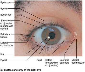
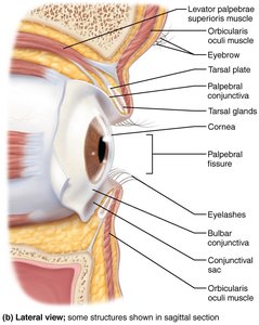
Structure of the Eyeball
The eyeball consists of three layers and internal chambers filled with fluids called humors.
Fibrous layer: Outermost, includes sclera and cornea.
Vascular layer: Middle, includes choroid, ciliary body, and iris.
Inner layer: Retina, contains photoreceptors.
Internal chambers: Anterior (aqueous humor) and posterior (vitreous humor) segments.
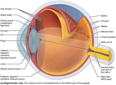
Fibrous Layer: Sclera and Cornea
The fibrous layer is dense, avascular connective tissue.
Sclera: Opaque, protects and shapes the eyeball, anchors muscles.
Cornea: Transparent, allows light entry and bends light, contains pain receptors.
Iris and Pupil
The iris is the colored part of the eye, controlling the size of the pupil and thus the amount of light entering the eye.
Pupil constriction: Sphincter pupillae contracts (parasympathetic control).
Pupil dilation: Dilator pupillae contracts (sympathetic control).
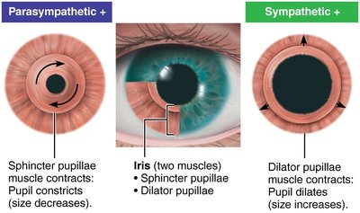
Retina: Structure and Function
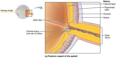
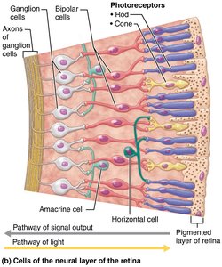
The retina originates as an outpocketing of the brain and contains millions of photoreceptor cells, neurons, and glial cells. It is a two-layered membrane:
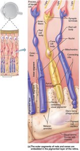
Outer pigmented layer: Absorbs light, stores vitamin A, phagocytizes cell fragments.
Inner neural layer: Contains photoreceptors (rods and cones), bipolar cells, and ganglion cells.
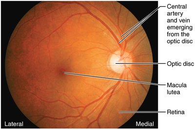
Optic disc: Site where optic nerve exits; lacks photoreceptors (blind spot).
Internal Chambers and Fluids
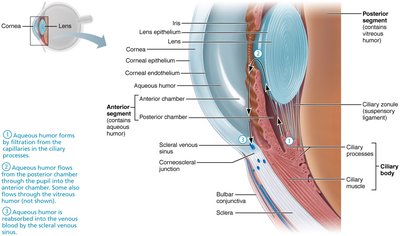
The lens and ciliary zonule separate the eye into two segments:
Posterior segment: Contains vitreous humor, supports lens and retina.
Anterior segment: Contains aqueous humor, supplies nutrients and removes wastes.
Lens
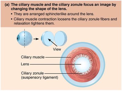
The lens is biconvex, transparent, flexible, and avascular. It changes shape to focus light on the retina. With age, the lens becomes more dense and less elastic.
Light and Optics
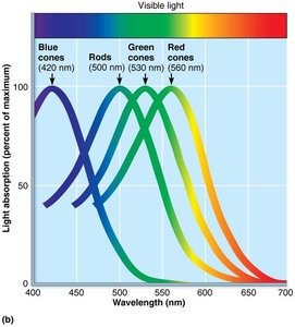
Light is composed of photons traveling in waves. The visible spectrum is perceived as different colors, depending on wavelength.
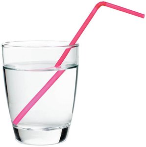
Refraction: Bending of light rays as they pass through different media.
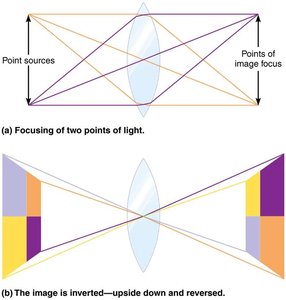
Lenses: Curved surfaces refract light to focus images.
Focusing Light on the Retina
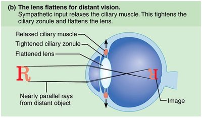
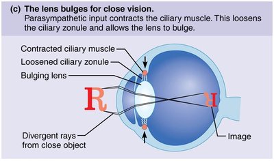
Light is refracted three times: entering the cornea, entering the lens, and leaving the lens. The cornea provides most of the refractory power, but the lens adjusts curvature for fine focusing.
Distant vision: Lens flattens; ciliary muscle relaxes.
Close vision: Lens bulges; ciliary muscle contracts.
Clinical Refraction Problems
Common refraction issues include:
Presbyopia: Loss of accommodation with age.
Myopia (nearsightedness): Eyeball too long; focal point in front of retina. Corrected with concave lens.
Hyperopia (farsightedness): Eyeball too short; focal point behind retina. Corrected with convex lens.
Astigmatism: Unequal curvatures; corrected with cylindrical lenses or laser.
Photoreceptors: Rods and Cones
Photoreceptors are modified neurons with cell body, synaptic terminal, and two segments. The outer segment contains visual pigments that change shape as they absorb light.
Rods: Sensitive to dim light, provide night and peripheral vision, converge for fuzzy images.
Cones: Require bright light, provide color and high-resolution vision, non-converging pathways.
Color Vision and Clinical Imbalances
Color blindness results from the absence of one or more cone pigments, most commonly red-green. It is inherited as an X-linked trait and more common in males.
Information Processing in the Retina
Photoreceptors and bipolar cells generate graded potentials. Light hyperpolarizes photoreceptors, reducing glutamate release, allowing bipolar cells to depolarize and activate ganglion cells, which generate action potentials sent to the brain.
Light and Dark Adaptation
Light adaptation: Rods and cones strongly stimulated; glare occurs; visual acuity improves as rods turn off and cones adapt.
Dark adaptation: Cones stop functioning; rod pigments regenerate; sensitivity increases over 20–30 minutes.
Visual Pathway to the Brain
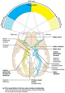
Axons of retinal ganglion cells form the optic nerve. Medial fibers cross at the optic chiasma, forming optic tracts that carry information to the thalamus and then to the primary visual cortex. Some fibers branch to midbrain for eye muscle control.
Depth Perception and Visual Processing
Both eyes view the same image from slightly different angles, and the visual cortex fuses these images for depth perception. Loss of one eye eliminates true depth perception.
Chemical Senses: Smell and Taste
Smell (Olfaction)
Smell is mediated by chemoreceptors in the olfactory epithelium, located in the roof of the nasal cavity. Olfactory neurons are bipolar and have cilia covered by mucus.
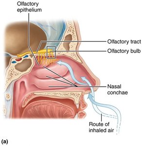
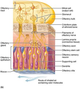
Olfactory neurons: Have stem cells that regenerate every 30–60 days.
Specificity: Humans have 350–400 smell genes, each encoding a unique receptor protein.
Pain and temperature receptors: Also present in nasal cavities.
Physiology of Smell
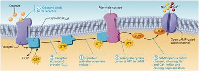
Odorants must be volatile and dissolve in olfactory epithelium fluid. They bind to receptor proteins, activating a G protein (Golf), which triggers cAMP synthesis and opens Na+ and Ca2+ channels, causing depolarization and adaptation.
Olfactory Pathway
Impulses travel from mitral cells via olfactory tracts to the olfactory cortex, thalamus, hypothalamus, amygdala, and limbic system, eliciting emotional responses to odors.
Taste (Gustation)
Taste buds are sensory organs located mainly on the tongue in papillae. Each taste bud contains gustatory epithelial cells and basal stem cells.
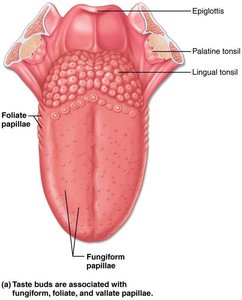
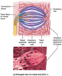
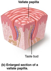
Gustatory epithelial cells: Have microvilli (gustatory hairs) that project into taste pores.
Basal epithelial cells: Divide every 7–10 days.
Basic Taste Sensations
Sweet: Sugars, saccharin, alcohol, some amino acids.
Sour: Hydrogen ions.
Salty: Metal ions.
Bitter: Alkaloids and some nonalkaloids.
Umami: Amino acids glutamate and aspartate.
Possible sixth taste: Long-chain fatty acids.
Physiology of Taste
To taste a chemical, it must be dissolved in saliva, diffuse into a taste pore, and contact gustatory hairs. Binding of tastant depolarizes the cell, releasing neurotransmitter and generating action potentials.
Gustatory Pathway
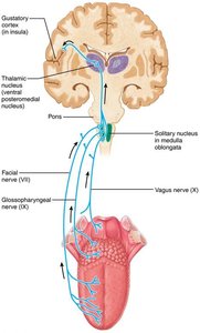
Taste impulses travel via the facial, glossopharyngeal, and vagus nerves to the medulla, thalamus, and gustatory cortex in the insula. The hypothalamus and limbic system are involved in taste appreciation.
Influence of Other Sensations on Taste
Taste is 80% smell. Thermoreceptors, mechanoreceptors, and nociceptors in the mouth enhance or detract from taste. Spicy foods excite pain receptors.
Hearing and Balance: The Ear
Structure of the Ear
The ear has three major areas: external, middle, and internal ear. The external and middle ear are involved in hearing, while the internal ear is involved in both hearing and equilibrium.
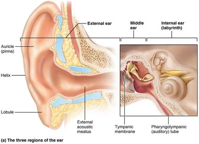
External ear: Auricle (pinna) and external acoustic meatus funnel sound waves.
Middle ear: Tympanic cavity, auditory ossicles (malleus, incus, stapes), and pharyngotympanic tube.
Internal ear: Bony and membranous labyrinth, vestibule, semicircular canals, cochlea.
Middle Ear
Auditory ossicles: Transmit vibratory motion from eardrum to oval window.
Pharyngotympanic tube: Equalizes pressure.
Internal Ear
Bony labyrinth: Filled with perilymph; includes vestibule, semicircular canals, cochlea.
Membranous labyrinth: Filled with endolymph; contains equilibrium and hearing receptors.
Hearing: Frequency and Amplitude
Frequency: Number of waves per second (Hz); determines pitch.
Amplitude: Height of waves; determines loudness (dB).
Transmission of Sound
Sound waves enter the external acoustic meatus, vibrate the tympanic membrane, and are amplified by the ossicles before reaching the oval window.
Sound Transduction
Movement of the basilar membrane deflects hair cells in the cochlea, opening ion channels and generating receptor potentials that trigger action potentials in the cochlear nerve.
The Auditory Pathway
Neural impulses travel from cochlear bipolar cells to the auditory cortex via the spiral ganglion, cochlear nuclei, superior olivary nucleus, lateral lemniscus, inferior colliculus, medial geniculate nucleus, and primary auditory cortex.
Equilibrium: Vestibular Apparatus
Vestibular apparatus: Includes maculae (static equilibrium) and crista ampullaris (dynamic equilibrium).
Maculae: Monitor head position and linear acceleration.
Crista ampullaris: Responds to rotational movements.
Summary
Vision
Light activates visual pigments in retinal photoreceptors, producing graded potentials. Visual information passes along the optic nerve to the thalamus and visual cortex for processing.
Taste
Chemicals dissolved in saliva activate gustatory hairs, producing graded potentials and neurotransmitter release. Afferent fibers travel via cranial nerves to the gustatory cortex.
Smell
Odorants dissolve in nasal fluids, bind to olfactory receptors, and activate neural pathways to the olfactory cortex and limbic system.
Hearing
Sound waves vibrate the eardrum and ossicles, press fluid in the internal ear, and stimulate hair cells, generating action potentials sent to the auditory cortex.
Balance
Equilibrium relies on input from the inner ear, eyes, and stretch receptors. Vestibular receptors monitor static and dynamic equilibrium, allowing for posture and spatial orientation.
Special Sense | Receptor Type | Location | Primary Function |
|---|---|---|---|
Vision | Photoreceptors (rods, cones) | Retina | Detect light, color, and movement |
Taste | Gustatory epithelial cells | Taste buds (tongue) | Detect dissolved chemicals |
Smell | Olfactory sensory neurons | Olfactory epithelium (nasal cavity) | Detect volatile chemicals |
Hearing | Hair cells | Cochlea (inner ear) | Detect sound waves |
Balance | Maculae, crista ampullaris | Vestibule, semicircular canals (inner ear) | Detect head position and movement |
References: Marieb, E. N., & Hoehn, K. (2023). Human anatomy and physiology (12th ed.). Pearson.
