 Back
BackSpinal Cord Anatomy and Reflexes: Structure, Function, and Clinical Relevance
Study Guide - Smart Notes
 Tailored notes based on your materials, expanded with key definitions, examples, and context.
Spinal Cord Anatomy
Gross Anatomy of the Spinal Cord
The spinal cord is a vital part of the central nervous system, extending from the brainstem to the lower back. It is divided into 31 segments, each associated with a pair of spinal nerves. These segments are classified as cervical (C1–C8), thoracic (T1–T12), lumbar (L1–L5), sacral (S1–S5), and coccygeal (Co1). Areas with increased gray matter, such as the cervical and lumbar enlargements, correspond to regions that control the limbs.
Cervical enlargement: Supplies nerves to the shoulder and upper limbs.
Lumbar enlargement: Supplies nerves to the pelvis and lower limbs.
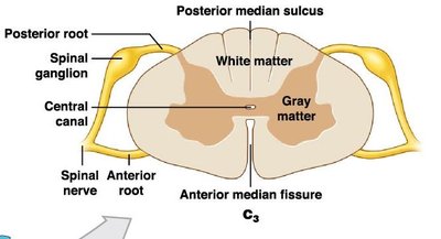
Spinal Cord Cross-Section
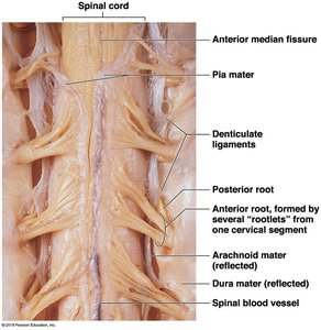
A cross-section of the spinal cord reveals two main types of tissue: gray matter (processing information) and white matter (transmitting information). Key anatomical landmarks include the anterior median fissure and posterior median sulcus, which help orient the structure.
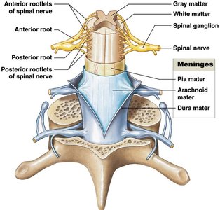
Spinal Nerve Roots and Ganglia
Each spinal segment has a pair of dorsal root ganglia containing the cell bodies of sensory (unipolar) neurons. The axons of these neurons form the dorsal roots, which join with ventral roots (containing motor neuron axons) to form mixed spinal nerves.
Dorsal root: Sensory axons only
Ventral root: Motor axons only
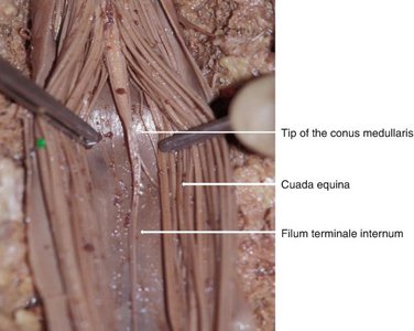
Base of the Spinal Cord
The conus medullaris is the tapered end of the spinal cord (L1–L2). The filum terminale is a fibrous strand extending from the conus medullaris, anchoring the cord. The cauda equina consists of long dorsal and ventral roots from L2–S5.
Anchoring the Spinal Cord
The spinal cord is anchored by the coccygeal ligament (prevents superior-inferior movement) and denticulate ligaments (prevent side-to-side movement).
Spinal Meninges
Structure and Function
The spinal cord is protected by three connective tissue layers called meninges:
Dura mater: Tough, outermost layer; separated from vertebrae by the epidural space (contains areolar tissue, blood vessels, and adipose tissue).
Arachnoid mater: Middle layer; delicate network of fibers. The subarachnoid space beneath it is filled with cerebrospinal fluid (CSF).
Pia mater: Innermost layer; firmly attached to the spinal cord surface.

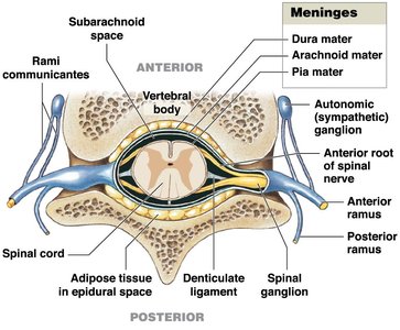
Lumbar Puncture (Spinal Tap)
A lumbar puncture is performed by inserting a needle into the subarachnoid space within the cauda equina region to collect CSF for diagnostic purposes.
Gray and White Matter Organization
Organization of Gray Matter
Gray matter in the spinal cord forms an H-shaped region and is divided into posterior (dorsal), lateral, and ventral (anterior) horns. The central canal runs through the center, containing CSF. The gray commissure consists of unmyelinated axons crossing from one side to the other.
Sensory nuclei: Located in the posterior horns; receive sensory input from the periphery.
Motor nuclei: Located in the anterior horns; send motor commands to effectors.
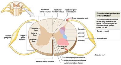
Organization of White Matter
White matter is organized into columns (posterior, lateral, anterior), each containing tracts of axons with similar functions. Ascending tracts carry sensory information to the brain, while descending tracts carry motor commands from the brain. The anterior white commissure contains myelinated axons crossing sides.

Functions of Gray and White Matter
Sensory information: Processed and carried on the dorsal (posterior) side; dorsal root and ganglion contain sensory axons and cell bodies.
Motor information: Processed and carried on the ventral (anterior) side; ventral root contains motor axons.
Spinal Nerves
Anatomy of Spinal Nerves
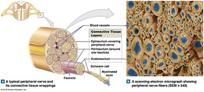
Spinal nerves are mixed nerves, containing both sensory and motor fibers. They are formed by the joining of dorsal and ventral roots and are covered by three connective tissue layers:
Epineurium: Surrounds the entire nerve
Perineurium: Divides nerve into fascicles (bundles)
Endoneurium: Surrounds individual axons
Dermatomes
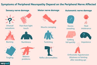
A dermatome is a region of skin supplied by a single pair of spinal nerves. Dermatomes are clinically important for diagnosing nerve injuries and neuropathies (regional losses of sensory/motor function due to trauma or compression).
Nerve Plexuses
Nerve plexuses are branching networks of intersecting nerves. The four major plexuses are cervical, brachial, lumbar, and sacral. These plexuses allow for redundancy and distribution of nerve fibers to limbs and body regions.
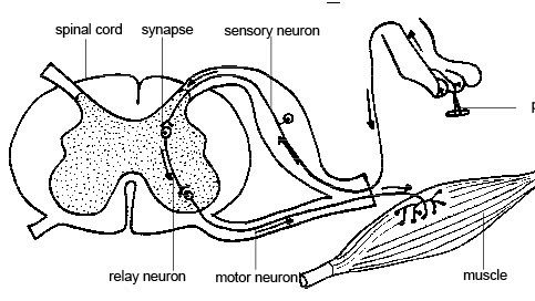
Reflexes and Reflex Arcs
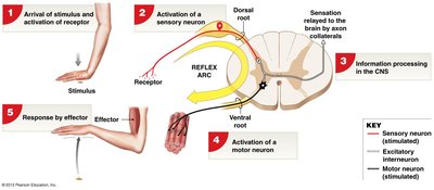
Reflex Arc
A reflex arc is the basic functional unit of the nervous system, beginning at a receptor and ending at a peripheral effector. Reflexes are typically rapid, automatic responses to stimuli and often involve negative feedback mechanisms.
Example: Withdrawal reflex (5 steps: receptor activation, sensory neuron activation, CNS processing, motor neuron activation, effector response)
Classification of Reflexes
Reflexes can be classified by development, response, complexity, and processing site:
Development | Response | Complexity | Processing Site |
|---|---|---|---|
Innate (genetically determined) | Somatic (skeletal muscle) | Monosynaptic (one synapse) | Spinal (spinal cord) |
Acquired (learned) | Visceral (smooth/cardiac muscle, glands) | Polysynaptic (multiple synapses) | Cranial (brain) |
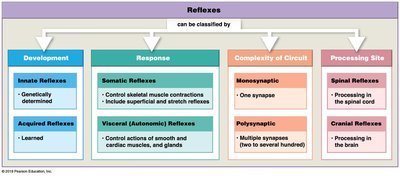
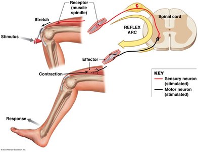
Monosynaptic Reflexes
Monosynaptic reflexes involve a direct connection between a sensory and a motor neuron, making them the fastest reflexes. The stretch reflex (e.g., patellar reflex) helps regulate muscle length and maintain posture.
Polysynaptic Reflexes
Polysynaptic reflexes involve multiple synapses and interneurons. The flexor reflex withdraws a limb from a painful stimulus, while reciprocal inhibition prevents antagonistic muscles from contracting. The crossed extensor reflex complements the flexor reflex by activating muscles on the opposite side of the body.


Additional info: Reflexes are essential for survival, allowing the body to respond rapidly to potentially harmful stimuli without conscious thought. Understanding the organization and function of the spinal cord is crucial for diagnosing and treating neurological disorders.
