 Back
BackSpinal Cord Anatomy and Reflexes: Structured Study Notes
Study Guide - Smart Notes
 Tailored notes based on your materials, expanded with key definitions, examples, and context.
Spinal Cord Anatomy
Gross Anatomy of the Spinal Cord
The spinal cord is a vital structure of the central nervous system, extending from the brainstem to the lower back. It is divided into 31 segments, each associated with a pair of spinal nerves:
Cervical nerves (C1–C8)
Thoracic nerves (T1–T12)
Lumbar nerves (L1–L5)
Sacral nerves (S1–S5)
Coccygeal nerve (Co1)
Enlargements occur in the cervical and lumbar regions due to increased gray matter for limb control.
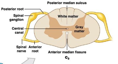
Spinal Cord Cross-Section
A cross-section of the spinal cord reveals two main types of tissue: gray matter (processing information) and white matter (transmitting information). Key anatomical landmarks include the anterior median fissure and posterior median sulcus.

Spinal Roots and Ganglia
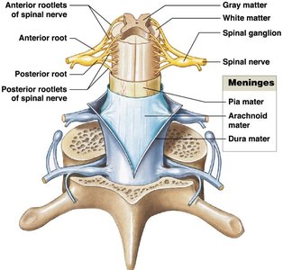
Each spinal segment has a pair of dorsal root ganglia containing sensory neuron cell bodies. The dorsal roots carry sensory axons, while ventral roots carry motor axons. These roots join to form mixed spinal nerves.
Base of the Spinal Cord
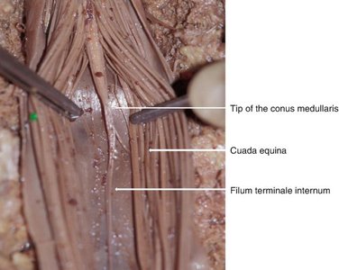
The spinal cord tapers at the conus medullaris (L1–L2). The filum terminale is a fibrous strand anchoring the cord, and the cauda equina consists of long dorsal and ventral roots below the conus medullaris.
Anchoring the Spinal Cord
The spinal cord is stabilized by the coccygeal ligament (prevents superior-inferior movement) and dentate ligaments (prevent side-to-side movement).
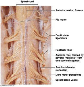
Spinal Meninges
Structure and Function
The meninges are three protective membranes covering the brain and spinal cord:
Dura mater: Tough, outermost layer; separated from vertebrae by the epidural space (contains areolar tissue, blood vessels, and adipose tissue).
Arachnoid mater: Middle, web-like layer; subarachnoid space beneath is filled with cerebrospinal fluid (CSF).
Pia mater: Thin, innermost layer, tightly bound to neural tissue.
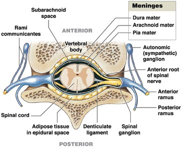
A lumbar puncture (spinal tap) is performed in the subarachnoid space within the cauda equina region to collect CSF.


Gray and White Matter Organization
Gray Matter
Gray matter forms an H-shaped core and is divided into posterior, lateral, and ventral horns. The central canal runs through the center, containing CSF. The gray commissure allows axons to cross sides.
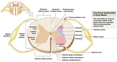
Nuclei are clusters of neuron cell bodies in the CNS:
Sensory nuclei: Located in dorsal horns; receive sensory input.
Motor nuclei: Located in ventral horns; send motor commands to effectors.

White Matter
White matter is organized into columns (posterior, lateral, anterior) containing tracts of axons:
Ascending tracts: Carry sensory information to the brain.
Descending tracts: Carry motor commands from the brain.
Anterior white commissure: Myelinated axons crossing sides.

Functions of Gray and White Matter
Sensory Information
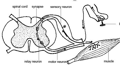
Sensory information enters the spinal cord via the dorsal root and is processed in the posterior horns. The dorsal root ganglion contains the cell bodies of sensory neurons.
Motor Information
Motor commands exit the spinal cord via the ventral root and are processed in the anterior horns. Motor neuron cell bodies are located in the ventral horn.
Spinal Nerves
Anatomy of Spinal Nerves
Spinal nerves are mixed nerves containing both sensory and motor fibers. They are formed by the joining of dorsal and ventral roots and are covered by three connective tissue layers:
Epineurium: Surrounds the entire nerve.
Perineurium: Surrounds bundles (fascicles) of axons; contains blood vessels.
Endoneurium: Surrounds individual axons; contains capillaries.
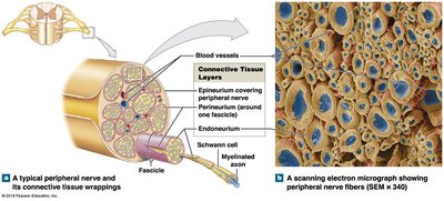
As spinal nerves branch into the periphery, they form smaller peripheral nerves.
Dermatomes and Nerve Plexuses
Dermatomes
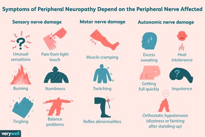
A dermatome is a region of skin supplied by a single pair of spinal nerves. Dermatomes are clinically important for diagnosing nerve injuries or neuropathies (regional sensory/motor loss due to trauma or compression).
Nerve Plexuses
A nerve plexus is a network of intersecting nerves. The four major plexuses are cervical, brachial, lumbar, and sacral.
Reflexes and Reflex Arcs
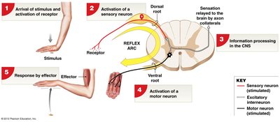
Reflex Arc
A reflex arc is the neural pathway that mediates a reflex, beginning at a receptor and ending at an effector. Reflexes typically involve negative feedback to maintain homeostasis.
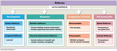
Classification of Reflexes
Reflexes can be classified by development, response, circuit complexity, and processing site:
Development | Response | Complexity of Circuit | Processing Site |
|---|---|---|---|
Innate (genetically determined) | Somatic (skeletal muscle) | Monosynaptic (one synapse) | Spinal (spinal cord) |
Acquired (learned) | Visceral (smooth/cardiac muscle, glands) | Polysynaptic (multiple synapses) | Cranial (brain) |
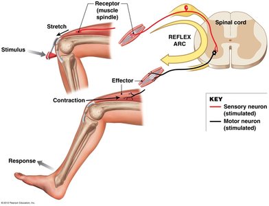
Monosynaptic Reflexes
The stretch reflex is a monosynaptic reflex that contracts a muscle in response to stretching, helping regulate muscle length and prevent overstretching. The patellar reflex is a classic example.
Polysynaptic Reflexes
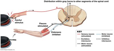
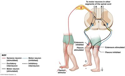
Polysynaptic reflexes involve multiple synapses and interneurons. The flexor reflex withdraws a limb from a painful stimulus, while reciprocal inhibition prevents antagonistic muscles from contracting simultaneously.
The crossed extensor reflex is a contralateral reflex arc that complements the flexor reflex by extending the opposite limb for balance.
Additional info: The spinal cord is essential for integrating sensory and motor information, and its organization allows for rapid reflex responses critical for survival and homeostasis.
