 Back
BackSpinal Cord and Spinal Nerves: Structure, Function, and Organization
Study Guide - Smart Notes
 Tailored notes based on your materials, expanded with key definitions, examples, and context.
Spinal Cord: Structure and Functions
Overview of Spinal Cord Functions
The spinal cord is a vital component of the central nervous system (CNS), serving as the main pathway for information connecting the brain and peripheral nervous system. It is responsible for conduction, neural integration, locomotion, and reflexes.
Conduction: Transmits sensory and motor signals between the body and brain.
Neural Integration: Processes and integrates information from various sources for appropriate responses.
Locomotion: Coordinates repetitive movements such as walking through central pattern generators.
Reflexes: Mediates rapid, involuntary responses to stimuli for protection and homeostasis.
Gross and Microscopic Anatomy of the Spinal Cord
External Structure and Regions
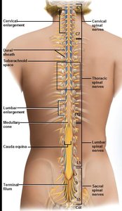
The spinal cord extends from the foramen magnum to the level of the first or second lumbar vertebra. It is divided into cervical, thoracic, lumbar, and sacral regions, with two enlargements (cervical and lumbar) for limb innervation. The cord tapers to the conus medullaris and continues as the cauda equina.
Cervical Enlargement: Supplies nerves to the upper limbs.
Lumbar Enlargement: Supplies nerves to the lower limbs.
Conus Medullaris: Tapered end of the spinal cord.
Cauda Equina: Bundle of nerve roots extending below the spinal cord.
Cross-Sectional Anatomy
A cross-section of the spinal cord reveals central gray matter (shaped like a butterfly) surrounded by white matter. The gray matter contains neuron cell bodies, while the white matter consists of myelinated axons organized into tracts.
Gray Matter: Contains dorsal (posterior) horns, ventral (anterior) horns, and lateral horns (in thoracic/lumbar regions).
White Matter: Organized into dorsal, lateral, and ventral columns (funiculi), each containing ascending and descending tracts.
Central Canal: Runs through the center, containing cerebrospinal fluid (CSF).
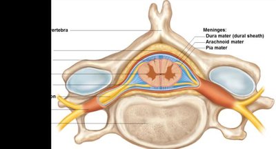
Spinal Meninges
The spinal cord is protected by three connective tissue membranes called meninges:
Dura Mater: Outermost, tough layer forming the dural sheath.
Arachnoid Mater: Middle, web-like layer.
Pia Mater: Innermost, delicate layer adhering to the cord surface.
Spinal Cord Tracts and Pathways
Ascending (Sensory) and Descending (Motor) Tracts
White matter tracts carry information up and down the spinal cord. Ascending tracts transmit sensory information to the brain, while descending tracts carry motor commands from the brain to effectors.
Ascending Tracts: Include the gracile fasciculus, cuneate fasciculus, spinothalamic, and spinocerebellar tracts.
Descending Tracts: Include corticospinal, tectospinal, reticulospinal, rubrospinal, and vestibulospinal tracts.
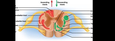
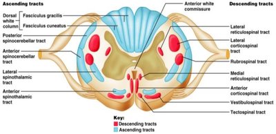
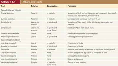
Table: Major Spinal Tracts
Tract | Column | Decussation | Functions |
|---|---|---|---|
Gracile fasciculus | Posterior | In medulla | Sensations of limb and trunk position and movement, deep touch, visceral pain, and vibration (below T6) |
Cuneate fasciculus | Posterior | In medulla | Same as gracile fasciculus (from level T6 up) |
Spinothalamic | Lateral and anterior | In spinal cord | Sensations of light touch, tickle, itch, temperature, pain, and pressure |
Spinoreticular | Lateral and anterior | In spinal cord (some fibers) | Sensation of pain from tissue injury |
Posterior spinocerebellar | Lateral | None | Feedback from muscles (proprioception) |
Anterior spinocerebellar | Lateral | In spinal cord | Same as posterior spinocerebellar |
Lateral corticospinal | Lateral | In medulla | Fine control of limbs |
Anterior corticospinal | Anterior | In spinal cord | Fine control of limbs |
Tectospinal | Anterior | In midbrain | Reflexive head turning in response to visual and auditory stimuli |
Reticulospinal | Lateral and anterior | None | Balance and posture; regulation of awareness of pain |
Rubrospinal | Lateral | In midbrain | Fine control of limbs |
Vestibulospinal | Anterior | Some fibers | Balance and posture |
Medial vestibulospinal | Anterior | Some fibers | Control of head position |
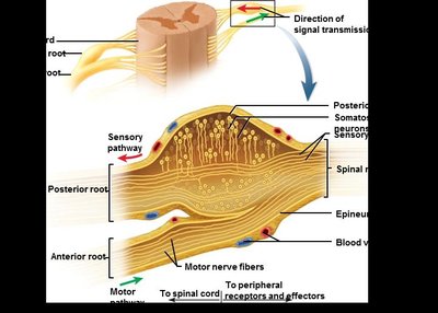
Spinal Nerves and Ganglia
General Structure of Spinal Nerves
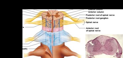
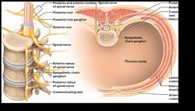
Spinal nerves are mixed nerves carrying both sensory (afferent) and motor (efferent) fibers. Each nerve is formed by the union of dorsal (sensory) and ventral (motor) roots. The nerves branch into rami that innervate specific body regions.
Rootlets: Small bundles of axons entering or leaving the spinal cord.
Posterior (Dorsal) Root: Contains sensory fibers and a dorsal root ganglion (cell bodies of sensory neurons).
Anterior (Ventral) Root: Contains motor fibers.
Spinal Nerve: Formed by the merging of dorsal and ventral roots; branches into dorsal and ventral rami.
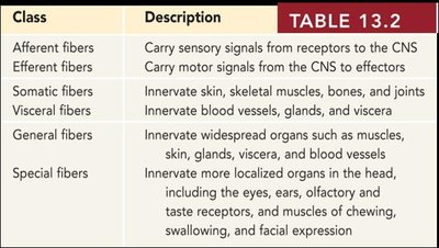
Classification of Nerve Fibers
Class | Description |
|---|---|
Afferent fibers | Carry sensory signals from receptors to the CNS |
Efferent fibers | Carry motor signals from the CNS to effectors |
Somatic fibers | Innervate skin, skeletal muscles, bones, and joints |
Visceral fibers | Innervate blood vessels, glands, and viscera |
General fibers | Innervate widespread organs such as muscles, skin, glands, viscera, and blood vessels |
Special fibers | Innervate more localized organs in the head, including the eyes, ears, olfactory and taste receptors, and muscles of chewing, swallowing, and facial expression |
Branches and Plexuses of Spinal Nerves
Proximal and Distal Branches
After emerging from the intervertebral foramen, each spinal nerve divides into several branches:
Meningeal Branch: Reenters the vertebral canal to innervate meninges and blood vessels.
Dorsal Ramus: Innervates muscles and skin of the back.
Ventral Ramus: Innervates anterior and lateral trunk, limbs, and forms nerve plexuses.
Communicating Rami: Connect spinal nerves to the sympathetic trunk (autonomic nervous system).
Nerve Plexuses
Nerve plexuses are networks of intersecting nerves formed by the ventral rami of spinal nerves. They provide redundancy and allow for complex innervation patterns.
Cervical Plexus (C1–C5): Innervates neck muscles and diaphragm.
Brachial Plexus (C5–T1): Innervates upper limb and shoulder.
Lumbar Plexus (L1–L4): Innervates anterior thigh and abdominal wall.
Sacral Plexus (L4–S4): Innervates posterior thigh, lower leg, and foot.
Coccygeal Plexus (S4–Co1): Innervates pelvic floor.
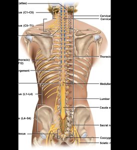
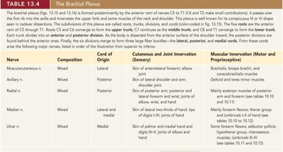
Table: The Brachial Plexus
Nerve | Composition | Cord of Origin | Cutaneous and Joint Innervation (Sensory) | Muscular Innervation (Motor and Proprioceptive) |
|---|---|---|---|---|
Musculocutaneous | Mixed | Lateral | Skin of anterolateral forearm, elbow joint | Brachialis, biceps brachii, coracobrachialis |
Axillary | Mixed | Posterior | Skin of lateral shoulder and arm, shoulder joint | Deltoid, teres minor |
Radial | Mixed | Posterior | Skin of posterior arm and forearm, posterolateral hand, joints of elbow, wrist, and hand | Mainly extensor muscles of posterior arm and forearm |
Median | Mixed | Lateral and medial | Skin of lateral two-thirds of hand, tips of digits I–IV, joints of hand | Mainly forearm flexors, some hand muscles |
Ulnar | Mixed | Medial | Skin of palmar and medial hand, digits III–V, joints of hand | Some forearm flexors, most intrinsic hand muscles |
Dermatomes and Spinal Nerve Distribution
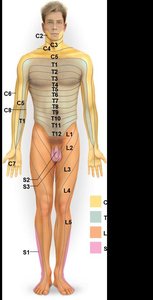
Dermatomes
A dermatome is an area of skin innervated by the sensory fibers of a single spinal nerve. Mapping dermatomes is clinically important for diagnosing nerve or spinal cord injuries.
Each spinal nerve (except C1) innervates a specific region of skin.
Dermatome maps help localize neurological lesions.
Summary of Key Concepts
The spinal cord is essential for conduction, integration, locomotion, and reflexes.
Its structure includes gray and white matter, protected by meninges.
Spinal nerves are mixed nerves with sensory and motor components, organized into plexuses and branches.
Dermatomes represent the cutaneous distribution of spinal nerves.
