 Back
BackSpinal Cord and Spinal Nerves: Structure, Function, and Protection
Study Guide - Smart Notes
 Tailored notes based on your materials, expanded with key definitions, examples, and context.
Spinal Cord and Spinal Nerves
Spinal Cord Protection
The spinal cord is protected by several anatomical structures that ensure its integrity and function. These include bony protection, connective tissue coverings, and specialized spaces filled with cerebrospinal fluid (CSF).
Bony Protection: The vertebral canal houses the spinal cord, shielding it from external injury.
Meninges: Three connective tissue layers surround the spinal cord and brain:
Dura mater: Outermost, dense irregular fibrous connective tissue; extends from foramen magnum to S2. Surrounded by the epidural space (fatty tissue).
Subdural space: Potential space between dura and arachnoid mater; may fill with blood after trauma (subdural hematoma).
Arachnoid mater: Middle, web-like layer; thinner than dura, made of fibrous and elastic tissue. The subarachnoid space between arachnoid and pia contains CSF.
Pia mater: Innermost, highly vascular, directly against spinal cord tissue. Denticulate ligaments (lateral extensions) stabilize the cord.
External Anatomy of the Spinal Cord
The spinal cord's external features are important for understanding its function and clinical relevance.
Length: Extends from foramen magnum to L1; about ¾” in diameter.
Conus medullaris: Tapered end of the cord.
Shape: Cylindrical, slightly flattened anteriorly and posteriorly.
Cervical and Lumbar Enlargements: Widenings where major nerve plexuses emerge.
Cauda equina: Nerve root fibers extending from lower cord.
Filum terminale: Extension of pia mater anchoring cord to coccyx.
Anterior median fissure: Deep indentation on anterior cord.
Posterior median sulcus: Lesser indentation on posterior cord.
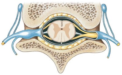
Internal Anatomy of the Spinal Cord
The internal structure of the spinal cord consists of gray and white matter, each with distinct functions and regions.
Gray Matter: Butterfly-shaped, contains unmyelinated cell bodies, neuroglia, and axons.
Posterior (Dorsal) horn: Receives sensory information.
Anterior (Ventral) horn: Contains motor neuron cell bodies (lower motor neurons).
Lateral horn: Origin of sympathetic and parasympathetic efferent nerves.
Gray commissure: Crossbar connecting right and left sides (association/interneurons).
Central canal: Runs length of cord, continuous with 4th ventricle, contains CSF.
White Matter: Surrounds gray matter, composed of myelinated axons.
Regions: Anterior, posterior, and lateral columns.

Specific Tracts of the White Matter
White matter contains ascending and descending tracts that transmit sensory and motor information.
Ascending Tracts: Carry sensory information to the brain.
Anterior Spinothalamic tract: Touch and pressure.
Lateral Spinothalamic tract: Pain and temperature.
Fasciculus cuneatus and gracilis: Discriminative touch, proprioception, vibration, stereognosis, and weight discrimination.
Descending Tracts: Carry motor information from the brain.
Anterior corticospinal tract: Controls trunk muscles.
Lateral corticospinal tract: Controls extremity movement.

Spinal Nerve Anatomy
Spinal nerves are essential for communication between the spinal cord and the body. There are 31 pairs, each with specific roots and branches.
Spinal Nerves:
8 cervical
12 thoracic
5 lumbar
5 sacral
1 coccygeal
Roots:
Posterior (Dorsal) root: Sensory fibers entering dorsal horn; dorsal root ganglion contains cell bodies of unipolar sensory neurons.
Anterior (Ventral) root: Motor fibers leaving ventral horn to periphery.

Branches of a Typical Spinal Nerve
After exiting the vertebral column, spinal nerves divide into several branches, each serving different regions and functions.
Posterior (Dorsal) ramus: Supplies muscles and skin of posterior trunk.
Anterior (Ventral) ramus: Supplies anterior/lateral trunk and extremities; forms plexuses.
Rami Communicantes: Contains visceral efferents for smooth muscle and glands.

Spinal Nerve Coverings
Spinal nerves are protected by three connective tissue layers:
Epineurium: Surrounds entire nerve; continuous with dura mater.
Perineurium: Surrounds groups of nerve fibers (fascicles).
Endoneurium: Surrounds individual nerve fibers.
Plexi and Nerves of the Body
Spinal nerves form plexuses that innervate specific regions and muscles.
Cervical Plexus (C1-C5): Skin and muscles of head, neck, upper shoulders.
Phrenic Nerve: Diaphragm.
Brachial Plexus (C5-T1): Skin and muscles of upper extremity.
Musculocutaneous Nerve: Biceps, Brachialis, Coracobrachialis.
Median Nerve: Pronators, flexors (except flexor carpi ulnaris), thenar muscles, first two lumbricales; sensory to anterior palm, thumb, lateral fingers.
Ulnar Nerve: Flexor carpi ulnaris, remaining hand muscles; sensory to medial hand, pinky, medial ring finger.
Axillary Nerve: Deltoid, Teres minor; sensory over deltoid and posterior arm.
Radial Nerve: Extensor muscles; sensory to posterior arm, forearm, hand.
Thoracic Nerves (T2-T12): Intercostal spaces, posterior trunk, upper abdominal muscles; sensory to skin of thorax.
Lumbar Plexus (L1-L4): Abdomen, genitals, anterior/medial thigh.
Femoral Nerve: Iliopsoas, Pectineus, Sartorius, Quadriceps.
Sacral Plexus (L4-S4): Buttock, perineum, posterior thigh, leg, foot.
Sciatic Nerve: Hamstrings, posterior adductor magnus.
Tibial Nerve: Posterior leg and foot muscles.
Superficial Peroneal Nerve: Lateral leg compartment.
Deep Peroneal Nerve: Anterior leg compartment.
Reflexes
Reflexes are rapid, automatic responses to environmental changes, mediated by a reflex arc.
Reflex: Fast, predictable, automatic response; inherent, not learned.
Reflex Arc: Five components:
Receptor: Senses environmental changes (e.g., free nerve endings, Pacinian corpuscles, muscle spindles).
Sensory neuron: Sends information from receptor to spinal cord.
Integrating center: Region in brain/spinal cord; relays information to motor neurons or other neurons.
Motor neuron: Sends information from ventral horn to effector.
Effector: Body part responding to motor neuron.
Additional info: Reflexes are clinically important for assessing nervous system function. Common examples include the patellar (knee-jerk) reflex and withdrawal reflex.
