Back
BackSpinal Cord and Spinal Nerves: Structure, Function, and Clinical Relevance
Study Guide - Smart Notes
 Tailored notes based on your materials, expanded with key definitions, examples, and context.
Spinal Cord and Spinal Nerves
Spinal Meninges and Associated Spaces
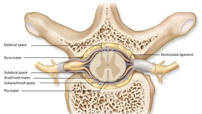
The spinal cord is protected by three connective tissue coverings known as the spinal meninges. These layers, along with associated spaces, provide structural support and protection for the central nervous system.
Dura mater: The tough, outermost layer.
Arachnoid mater: The middle, web-like layer.
Pia mater: The delicate, innermost layer that adheres to the spinal cord.
Denticulate ligaments: Extensions of the pia mater that anchor the spinal cord to the dura mater.
Epidural space: Located between the dura mater and the vertebral wall; contains fat and blood vessels.
Subdural space: Between the dura mater and arachnoid mater; contains a thin film of fluid.
Subarachnoid space: Between the arachnoid mater and pia mater; filled with cerebrospinal fluid (CSF).
Epidural Anesthesia and Lumbar Puncture
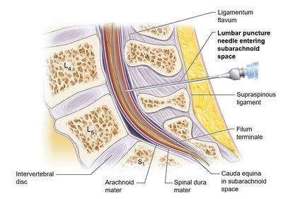
Medical procedures involving the spinal meninges include epidural anesthesia and lumbar puncture. Epidural anesthesia is administered into the epidural space to block pain, while lumbar puncture involves inserting a needle into the subarachnoid space to collect CSF for diagnostic purposes.
Epidural: Used for pain management, especially during childbirth.
Lumbar puncture: Used to diagnose infections, hemorrhages, and other CNS disorders.
Spinal Cord Anatomy and Segmentation
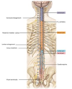
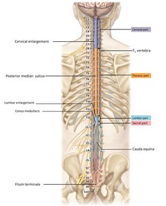
The spinal cord extends from the foramen magnum to the level of the second lumbar vertebra (L2). It is housed within the vertebral foramen and is divided into 31 segments, each giving rise to a pair of spinal nerves.
31 segments: Each segment corresponds to a pair of spinal nerves.
Spinal nerves: Mixed nerves carrying sensory and motor information.
External Anatomy of the Spinal Cord
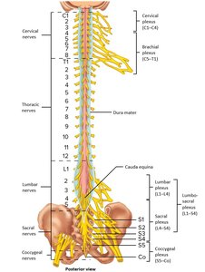
The external anatomy of the spinal cord includes regions of enlargement and terminal structures. These features are important for understanding nerve distribution and clinical landmarks.
Cervical enlargement: Supplies nerves to the upper limbs.
Lumbar enlargement: Supplies nerves to the lower limbs.
Conus medullaris: The tapered end of the spinal cord.
Cauda equina: A bundle of spinal nerves below the conus medullaris.
Filum terminale: A fibrous extension anchoring the spinal cord to the coccyx.
Internal Structure of the Spinal Cord
Gray Matter Organization
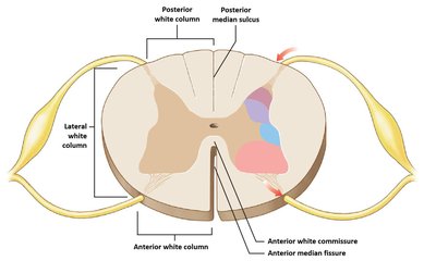
The spinal cord's gray matter is centrally located and organized into horns. Each horn contains nuclei responsible for processing different types of information.
Posterior (dorsal) horn: Contains sensory nuclei (somatic and visceral).
Anterior (ventral) horn: Contains motor nuclei (somatic).
Lateral horn: Contains visceral motor nuclei (present in thoracic and upper lumbar regions).
Central canal: Contains cerebrospinal fluid.
Gray commissures: Connect the two sides of gray matter.
White Matter Organization and Nerve Tracts
White matter surrounds the gray matter and is organized into columns (funiculi) containing myelinated axons. These columns house ascending (sensory) and descending (motor) nerve tracts.
Anterior, lateral, and posterior columns: Contain bundles of axons (fasciculi).
Ascending tracts: Carry sensory information to the brain.
Descending tracts: Carry motor commands from the brain.
Spinal Nerves and Their Structure
Spinal Roots and Ganglia
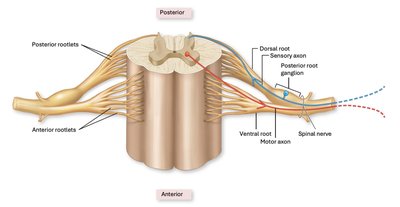
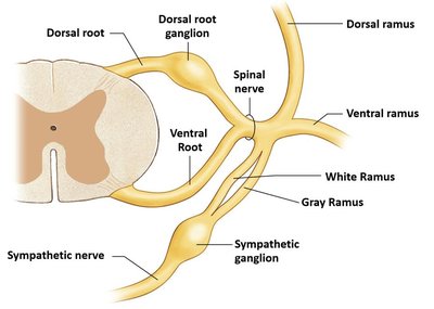
Each spinal nerve is formed by the union of a dorsal (sensory) root and a ventral (motor) root. The dorsal root contains a ganglion housing sensory neuron cell bodies.
Dorsal root: Carries sensory information to the spinal cord.
Dorsal root ganglion: Contains cell bodies of sensory neurons.
Ventral root: Carries motor information away from the spinal cord.
Spinal Nerve Structure
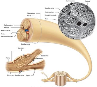
Spinal nerves are cable-like bundles of axons, organized into compartments by connective tissue layers. These layers provide protection and facilitate nerve function.
Epineurium: Outermost layer surrounding the entire nerve.
Perineurium: Surrounds bundles of axons (fascicles).
Endoneurium: Surrounds individual axons.
Myelin sheath: If present, located deep to the endoneurium.
Branches of Spinal Nerves
After exiting the vertebral column, spinal nerves branch into rami. These branches distribute sensory and motor fibers to different regions of the body.
Dorsal ramus: Supplies the muscles and skin of the back.
Ventral ramus: Supplies the limbs and anterior trunk.
White ramus: Contains myelinated preganglionic neurons.
Gray ramus: Contains unmyelinated postganglionic neurons.
Dermatomes and Nerve Plexuses
Dermatome Map and Peripheral Neuropathies
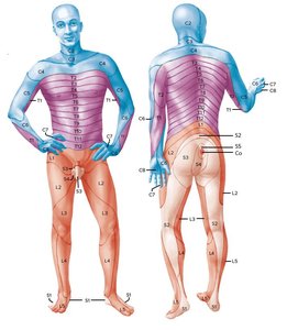
A dermatome is an area of skin supplied by a single spinal nerve. Understanding dermatomes is crucial for diagnosing nerve injuries and regional losses of sensory or motor function (peripheral neuropathies).
Dermatome: Skin region innervated by a specific spinal nerve.
Peripheral neuropathies: Regional losses of sensory and motor function due to nerve damage.
Spinal Nerves and Nerve Plexuses
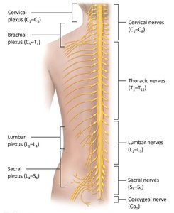
There are 31 pairs of spinal nerves, grouped by region. Many spinal nerves form networks called nerve plexuses, which allow for complex innervation patterns.
Cervical: C1–C8
Thoracic: T1–T12
Lumbar: L1–L5
Sacral: S1–S5
Coccygeal: Co1
Nerve plexus: Network of interwoven nerve fibers (cervical, brachial, lumbar, sacral).
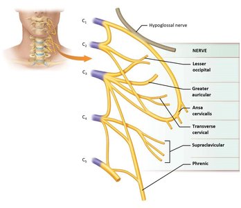
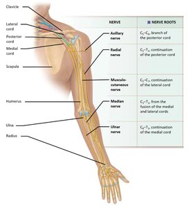
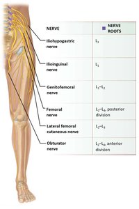
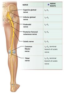
Cervical, Brachial, Lumbar, and Sacral Plexuses
Each plexus is formed by ventral rami of specific spinal nerves and innervates distinct regions of the body.
Cervical plexus: Ventral rami C1–C5; innervates neck and diaphragm.
Brachial plexus: Ventral rami C5–T1; innervates upper limb.
Lumbar plexus: Ventral rami L1–L4; innervates anterior and medial thigh.
Sacral plexus: Ventral rami L4–S4; innervates posterior thigh, leg, and foot.
Reflexes and Spinal Cord Disorders
Reflexes and Reflex Arcs
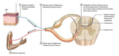
A reflex is a rapid, automatic response to a stimulus, mediated by a reflex arc. Reflexes are essential for maintaining homeostasis and protecting the body from harm.
Reflex arc: The neural pathway involved in a reflex.
Components: Sensory receptor, sensory neuron, integration center, motor neuron, effector.
Classification of Reflexes
Reflexes can be classified based on their development, effector type, processing site, and number of synapses.
Time of development: Innate (present at birth) or acquired (learned).
Type of effector: Somatic (skeletal muscle) or autonomic (smooth muscle, glands).
Processing site: Cranial (brain) or spinal (spinal cord).
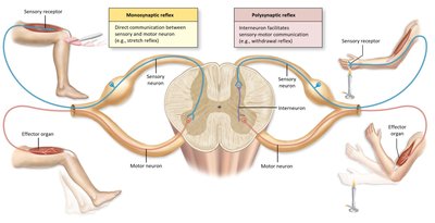
Number of synapses: Monosynaptic (one synapse) or polysynaptic (multiple synapses).
Stretch and Withdrawal Reflexes
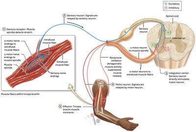
The stretch reflex causes muscle contraction in response to stretching, while the withdrawal reflex removes a body part from painful stimuli. Both are critical for motor control and protection.
Stretch reflex: Maintains muscle tone and posture.
Withdrawal reflex: Protects from injury by rapidly moving away from harmful stimuli.
Traumatic Injuries and Spinal Cord Disorders
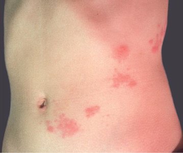
Traumatic injuries to the spinal cord can result in loss of function below the site of injury. Common disorders include paraplegia (loss of function in lower limbs), quadriplegia (loss of function in all limbs), shingles (viral infection affecting dermatomes), and polio (viral infection causing motor neuron damage).
Paraplegia: Paralysis of lower limbs.
Quadriplegia: Paralysis of all four limbs.
Shingles: Caused by reactivation of the varicella-zoster virus; affects specific dermatomes.
Polio: Caused by poliovirus; damages motor neurons.