Back
BackSpinal Cord and Spinal Nerves: Structure, Function, and Organization
Study Guide - Smart Notes
 Tailored notes based on your materials, expanded with key definitions, examples, and context.
Spinal Cord: Gross Anatomy
General Structure and Location
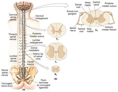
The spinal cord is a major component of the central nervous system, enclosed within the vertebral canal of the spinal column. It extends from the foramen magnum at the base of the skull to the level of the first or second lumbar vertebra (L1 or L2) in adults.
Cervical enlargement: Region where nerves serving the upper limbs arise.
Lumbar enlargement: Region where nerves serving the lower limbs arise.
Conus medullaris: Tapered end of the spinal cord.
Cauda equina: Collection of spinal nerves at the end of the spinal cord resembling a horse's tail.
Spinal Cord Segmentation
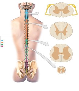
The spinal cord is divided into regions corresponding to the vertebral column: cervical, thoracic, lumbar, sacral, and coccygeal. There are 31 pairs of spinal nerves emerging from these regions.
8 pairs of cervical nerves (C1–C8)
12 pairs of thoracic nerves (T1–T12)
5 pairs of lumbar nerves (L1–L5)
5 pairs of sacral nerves (S1–S5)
1 pair of coccygeal nerves (Co1)

Spinal Cord Meninges
Protective Layers
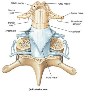
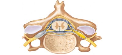
The spinal cord is protected by three connective tissue membranes called meninges:
Dura mater: Outermost layer; tough and fibrous.
Epidural space: Between vertebra and dura, filled with fat and blood vessels; site for epidural anesthesia.
Subdural space: Between dura and arachnoid.
Arachnoid mater: Middle layer; delicate and web-like.
Subarachnoid space: Between arachnoid and pia, filled with cerebrospinal fluid (CSF).
Pia mater: Innermost layer; clings tightly to the spinal cord.
Denticulate ligaments: Saw tooth-like shelves of pia mater securing the cord to dura.
Filum terminale: Fibrous extension of pia mater from conus medullaris to coccyx.

Clinical Application: Lumbar Tap
Lumbar tap: Removal of CSF from the vertebral canal below L3 for analysis or infection detection.
No danger to the spinal cord, which ends at L1–L2 in adults.
Internal Structure of the Spinal Cord
Gray Matter and White Matter
The spinal cord consists of two main tissue types:
White matter: Outer portion; contains myelinated axons organized into columns (funiculi) and tracts.
Gray matter: Inner portion; butterfly-shaped; contains cell bodies, unmyelinated axons, dendrites, and neuroglia.
Gray Matter Regions
Posterior (dorsal) horns: Somatic and visceral sensory fibers; interneurons.
Anterior (ventral) horns: Cell bodies of somatic motor neurons; axons exit via ventral root.
Lateral gray horns: Visceral motor neurons; present only in thoracic and lumbar regions.
Gray commissure: Axons crossing from one side to the other.
Central canal: Lined with ependymal cells; filled with CSF.
White Matter Regions
Posterior funiculi: Located near the posterior median sulcus.
Anterior funiculi: Located near the anterior median fissure.
Lateral funiculi: Located on each side.
Key Vocabulary
Nuclei: Collection of cell bodies of neurons in the CNS.
Ganglion: Collection of cell bodies of neurons in the PNS.
Spinal Nerves and Their Organization
Formation and Branching
Spinal nerves are formed by the fusion of ventral and dorsal roots. Each spinal nerve quickly divides into two rami:
Dorsal ramus: Sensory and motor innervation to skin and muscles of the back.
Ventral ramus:
T2–T12 form intercostal nerves (innervate intercostal spaces, anterior/lateral trunk).
Other ventral rami form plexuses (innervate limbs).
Rami communicantes: Communicate with sympathetic chain ganglia (ANS innervation).
Nerve Plexuses
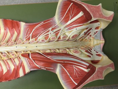
Complex interwoven networks formed by ventral rami of spinal nerves. Four major plexuses:
Cervical plexus: C1–C5; innervates head, neck, diaphragm (phrenic nerve).
Brachial plexus: C5–T1; innervates arm muscles and skin (axillary, musculocutaneous, radial, median, ulnar nerves).
Lumbar plexus: T12–L5; serves lower limbs (femoral, obturator nerves).
Sacral plexus: L5–S5; serves lower limbs (sciatic nerve, tibial, common fibular nerves).
Major Peripheral Nerves and Their Innervation
Nerve | Plexus | Innervation |
|---|---|---|
Phrenic | Cervical | Diaphragm for respiration |
Axillary | Brachial | Shoulder muscles and skin |
Musculocutaneous | Brachial | Arm and forearm muscles and skin |
Radial | Brachial | Forearm muscles and skin |
Median | Brachial | Forearm muscles and skin |
Ulnar | Brachial | Forearm muscles and skin |
Femoral | Lumbar | Lower abdomen, anterior and medial thigh muscles and skin |
Obturator | Lumbar | Hip and medial thigh muscles and skin |
Sciatic | Sacral | Lower trunk, posterior thigh muscles and skin |
Tibial | Sacral (branch of sciatic) | Posterior leg and foot muscles and skin |
Common fibular | Sacral (branch of sciatic) | Lateral leg and foot muscles and skin |

Summary Table: Spinal Cord Regions and Nerves
Region | Number of Nerves | Major Plexus |
|---|---|---|
Cervical | 8 | Cervical, Brachial |
Thoracic | 12 | None (intercostal nerves) |
Lumbar | 5 | Lumbar |
Sacral | 5 | Sacral |
Coccygeal | 1 | None |
Key Concepts and Applications
Spinal cord anatomy is essential for understanding nervous system function and clinical procedures (e.g., lumbar tap).
Spinal nerves provide sensory and motor innervation to the body, organized by region and plexus.
Meninges protect the spinal cord and facilitate safe access for medical procedures.
Plexuses allow for complex innervation patterns, especially in the limbs.
Example: Sciatica
Sciatica: Stabbing pain radiating along the sciatic nerve, often due to spinal disc herniation.
Example: Phrenic Nerve
Phrenic nerve: Innervates the diaphragm, crucial for breathing.
Example: Lumbar Tap
Lumbar tap: Removal of CSF below L3, used for diagnostic purposes.