Back
BackSpinal Nerves and Plexuses Study Guide – Step-by-Step Guidance
Study Guide - Smart Notes
 Tailored notes based on your materials, expanded with key definitions, examples, and context.
Q1. What are the nerves that make up the: Cervical Plexus, Brachial Plexus, Lumbar Plexus, Sacral Plexus?
Background
Topic: Spinal Nerve Plexuses
This question tests your knowledge of the major nerve plexuses and the specific nerves that arise from each. Understanding these plexuses is essential for mapping nerve supply to different regions of the body.
Key Terms:
Plexus: A network of intersecting nerves.
Cervical, Brachial, Lumbar, Sacral: The four main plexuses formed by spinal nerves.
Step-by-Step Guidance
Recall the spinal nerve roots that contribute to each plexus (e.g., cervical plexus: C1–C5).
List the major nerves that arise from each plexus (e.g., phrenic nerve from cervical plexus).
Review diagrams or charts showing the branching of nerves from each plexus.
Compare the functions and regions supplied by each nerve within the plexus.
Try solving on your own before revealing the answer!
Q2. Match the nerves with the appropriate plexus.
Background
Topic: Nerve-Plexus Associations
This question assesses your ability to correctly associate specific nerves with their originating plexus.
Key Terms:
Common Fibular, Obturator, Phrenic, Radial, Musculocutaneous, Femoral, Ulnar, Sciatic, Inferior Gluteal: Major peripheral nerves.
Plexus: Cervical, Brachial, Lumbar, Sacral.
Step-by-Step Guidance
Review the anatomical location and function of each nerve listed.
Recall which plexus supplies which region (e.g., brachial plexus supplies the upper limb).
Match each nerve to its corresponding plexus based on its region and function.
Double-check your matches using a labeled diagram of the plexuses.
Try solving on your own before revealing the answer!
Q3. The lateral cord of the brachial plexus is associated with which nerve roots?
Background
Topic: Brachial Plexus Anatomy
This question tests your understanding of the organization of the brachial plexus, specifically the nerve roots that form the lateral cord.
Key Terms:
Lateral cord: One of the three cords of the brachial plexus.
Nerve roots: The spinal nerves (C5–T1) that contribute to the plexus.
Step-by-Step Guidance
Recall the structure of the brachial plexus: roots, trunks, divisions, cords.
Identify which roots contribute to the lateral cord (typically from the anterior divisions of the superior and middle trunks).
Review diagrams to confirm which nerve roots are involved.
Try solving on your own before revealing the answer!
Q4. The middle trunk of the brachial plexus is associated with which cords?
Background
Topic: Brachial Plexus Trunks and Cords
This question focuses on the relationship between the trunks and cords of the brachial plexus.
Key Terms:
Middle trunk: Formed by the C7 nerve root.
Cords: Lateral, posterior, medial.
Step-by-Step Guidance
Recall how the trunks divide into anterior and posterior divisions.
Determine which divisions from the middle trunk contribute to which cords.
Use a diagram to visualize the connections.
Try solving on your own before revealing the answer!
Q5. The Ulnar nerve is associated with which nerve roots, trunks, and cords?
Background
Topic: Ulnar Nerve Anatomy
This question tests your ability to trace the origin of the ulnar nerve through the brachial plexus.
Key Terms:
Ulnar nerve: Major nerve of the forearm and hand.
Nerve roots, trunks, cords: Components of the brachial plexus.
Step-by-Step Guidance
Identify the nerve roots that contribute to the ulnar nerve (typically C8 and T1).
Determine which trunk(s) these roots form (inferior trunk).
Trace the divisions and cords to see which cord gives rise to the ulnar nerve (medial cord).
Try solving on your own before revealing the answer!
Q6. List the nerves associated with each cord: Lateral cord, Posterior cord, Medial cord.
Background
Topic: Brachial Plexus Cord Nerves
This question tests your knowledge of which nerves arise from each cord of the brachial plexus.
Key Terms:
Lateral, posterior, medial cords: Major divisions of the brachial plexus.
Musculocutaneous, axillary, radial, median, ulnar: Major nerves associated with these cords.
Step-by-Step Guidance
Recall the nerves that branch from each cord (e.g., musculocutaneous from lateral cord).
Use a diagram to visualize the branching pattern.
List the nerves for each cord, stopping before the final answer.
Try solving on your own before revealing the answer!
Q7. Which two of the above cords are part of the anterior division?
Background
Topic: Brachial Plexus Divisions
This question tests your understanding of the divisions of the brachial plexus and which cords are formed from the anterior divisions.
Key Terms:
Anterior division: Part of the brachial plexus that forms certain cords.
Lateral and medial cords: Typically formed from anterior divisions.
Step-by-Step Guidance
Recall the structure of the brachial plexus: roots, trunks, divisions, cords.
Identify which cords are formed from the anterior divisions.
Try solving on your own before revealing the answer!
Q8–10. Which cord does the axillary, median, and radial nerve derive from?
Background
Topic: Brachial Plexus Cord-Nerve Relationships
These questions test your ability to identify which cord gives rise to each major nerve of the upper limb.
Key Terms:
Axillary, median, radial nerves: Major nerves of the upper limb.
Lateral, posterior, medial cords: Brachial plexus divisions.
Step-by-Step Guidance
Review the branching pattern of the brachial plexus cords.
Identify which cord gives rise to each nerve using a diagram.
Try solving on your own before revealing the answer!
Q12. List two muscles supplied by the cervical plexus.
Background
Topic: Cervical Plexus Muscle Supply
This question tests your knowledge of the muscles innervated by nerves from the cervical plexus.
Key Terms:
Cervical plexus: Network of nerves supplying neck muscles.
Phrenic nerve: Supplies the diaphragm.
Step-by-Step Guidance
Recall the major nerves of the cervical plexus and their target muscles.
List two muscles innervated by these nerves.
Try solving on your own before revealing the answer!
Q13–16. Which nerve roots give rise to the lateral femoral cutaneous, sciatic, tibial, and femoral nerves?
Background
Topic: Nerve Root Origins
These questions test your knowledge of the specific spinal nerve roots that form major peripheral nerves.
Key Terms:
Lateral femoral cutaneous, sciatic, tibial, femoral nerves: Major nerves of the lower limb.
Nerve roots: Spinal segments contributing to each nerve.
Step-by-Step Guidance
Recall the spinal segments associated with each nerve.
Use a diagram or chart to confirm the nerve root origins.
Try solving on your own before revealing the answer!
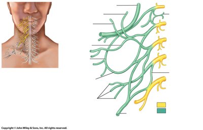
Q17. Label the illustration above using the following list: C1, C2, C3, C4, C5, to brachial plexus, Phrenic nerve, Hypoglossal nerve (XII), Supraclavicular Nerve, Lesser occipital nerve, Great auricular nerve, Transverse cervical nerve
Background
Topic: Cervical Plexus and Associated Nerves
This question tests your ability to identify and label the nerves and roots in a diagram of the cervical plexus.
Key Terms:
Cervical nerve roots: C1–C5.
Phrenic, hypoglossal, supraclavicular, lesser occipital, great auricular, transverse cervical nerves.
Step-by-Step Guidance
Review the diagram and locate each nerve/root listed.
Label each structure based on its anatomical position.
Try solving on your own before revealing the answer!
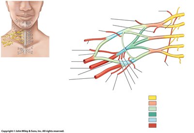
Q18. Label the illustration above using the following list: C5-T1 nerve roots, Superior, middle and inferior trunks, Lateral, posterior, and medial cords, Musculocutaneous nerve, Axillary nerve, Median nerve, Radial nerve, Ulnar nerve
Background
Topic: Brachial Plexus Anatomy
This question tests your ability to identify and label the components of the brachial plexus in a diagram.
Key Terms:
C5–T1 nerve roots, trunks, cords, major nerves of the upper limb.
Step-by-Step Guidance
Review the diagram and locate each structure listed.
Label each component based on its anatomical position and branching pattern.
Try solving on your own before revealing the answer!
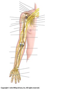
Q19. Label the illustration above using the following list: C5-T1 nerve roots, scapula, Ulnar nerve, humerus, radius, ulna, Ulnar nerve, Digital branch of median and ulnar nerves, Superficial branch of ulnar nerve, Radial nerve (twice & superficial and deep branches), Median nerve (twice), Musculocutaneous nerve, medial cord, posterior cord, lateral cord, Axillary nerve, Lateral pectoral nerve, Superior, middle, and inferior trunks
Background
Topic: Upper Limb Nerve Anatomy
This question tests your ability to identify and label the nerves and bones of the upper limb in a diagram.
Key Terms:
Upper limb nerves and bones: C5–T1 roots, scapula, humerus, radius, ulna, major nerves.
Step-by-Step Guidance
Review the diagram and locate each structure listed.
Label each nerve and bone based on its anatomical position.
Try solving on your own before revealing the answer!
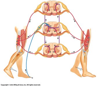
Q20. Practice labeling the arrows above.
Background
Topic: Spinal Reflex Pathways
This question tests your ability to identify and label the components of a spinal reflex pathway in a diagram.
Key Terms:
Spinal cord, sensory and motor neurons, reflex arc.
Step-by-Step Guidance
Review the diagram and identify the arrows representing sensory input, motor output, and interneuron connections.
Label each pathway based on its function in the reflex arc.
Try solving on your own before revealing the answer!