 Back
Spinal Nerves, Nerve Plexuses, and Peripheral Nerve Distribution
Study Guide - Smart Notes
 Tailored notes based on your materials, expanded with key definitions, examples, and context.
Spinal Nerves and Peripheral Nerves
Anatomy of Spinal Nerves
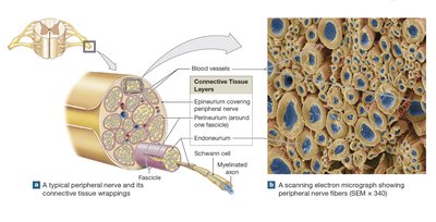
Spinal nerves are critical components of the peripheral nervous system, connecting the spinal cord to body tissues and organs. Each spinal nerve is surrounded by three connective tissue layers that provide structure and protection, similar to the organization seen in skeletal muscles.
Epineurium: The outermost layer, composed of dense collagen fibers, encases the entire nerve.
Perineurium: The middle layer, which divides the nerve into bundles called fascicles.
Endoneurium: The innermost layer, surrounding individual axons within each fascicle.
Blood Supply: Arteries and veins penetrate the epineurium and branch within the perineurium and endoneurium, supplying nutrients to axons and Schwann cells.
Peripheral nerves are extensions of spinal nerves, maintaining the same connective tissue sheaths. Recovery from peripheral nerve injury depends on the extent of damage; axons may regrow if alignment is preserved, but complete nerve damage often results in incomplete repair.
Peripheral Distribution and Function of Spinal Nerves
Each spinal nerve innervates a specific region of the skin, known as a dermatome. Dermatomes are clinically significant for diagnosing nerve or spinal cord injuries, as loss of sensation in a dermatome can indicate the location of damage.
Dermatome: A bilateral region of skin supplied by a single pair of spinal nerves. Adjacent dermatomes overlap slightly.
Peripheral Neuropathies: Regional losses of sensory and motor function, often due to trauma or compression (e.g., limb "falling asleep").
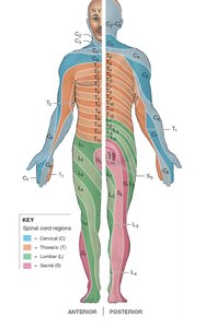
Mapping dermatomes helps localize spinal cord injuries, especially when combined with motor function assessment.
Clinical Note: Shingles
Shingles is caused by the varicella-zoster virus, which remains dormant in neurons after chickenpox. Reactivation leads to a painful rash and blisters along the affected dermatome. Treatment involves antiviral drugs, and vaccination is recommended for adults over 50.
Nerve Plexuses
Overview of Nerve Plexuses
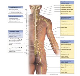
Nerve plexuses are complex networks formed by the blending of anterior rami of adjacent spinal nerves. These plexuses provide both sensory and motor innervation to the limbs and certain body regions. Only anterior rami form plexuses; posterior rami do not.
Cervical Plexus (C1–C5): Innervates neck muscles and diaphragm.
Brachial Plexus (C5–T1): Innervates pectoral girdle and upper limb.
Lumbar Plexus (T12–L4): Innervates pelvic girdle and lower limb.
Sacral Plexus (L4–S4): Innervates pelvic girdle and lower limb.
The Cervical Plexus
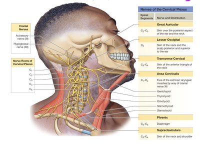
The cervical plexus is formed by the anterior rami of spinal nerves C1–C5. It innervates muscles of the neck and the diaphragm. The phrenic nerve is the major nerve, supplying the diaphragm and playing a vital role in breathing. Other branches serve the skin of the neck and upper chest.
Mnemonic: "C3, C4, C5 keep the diaphragm alive." (Phrenic nerve roots)
The Brachial Plexus
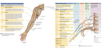
The brachial plexus is formed by the anterior rami of C5–T1 (sometimes C4 and T2). It innervates the pectoral girdle and upper limb. The plexus forms trunks (superior, middle, inferior) and cords (lateral, medial, posterior) before branching into peripheral nerves.
Major Nerves: Musculocutaneous, median, ulnar, axillary, and radial nerves.
Clinical Note: Carpal tunnel syndrome involves compression of nerves at the wrist, affecting sensation in the hand.
The Lumbar and Sacral Plexuses
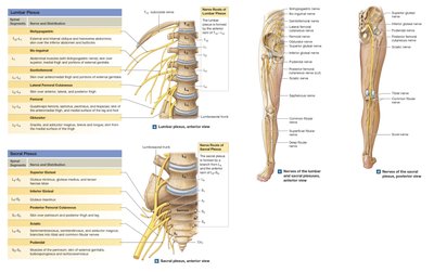
The lumbar and sacral plexuses innervate the pelvic girdle and lower limbs. The lumbar plexus (T12–L4) gives rise to the genitofemoral, lateral femoral cutaneous, and femoral nerves. The sacral plexus (L4–S4) gives rise to the sciatic and pudendal nerves. The sciatic nerve is the largest nerve in the body and divides into the common fibular and tibial nerves near the knee.
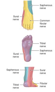
Sural Nerve: A sensory nerve of the lateral foot, often used for nerve grafts.
Clinical Note: Mapping sensory nerves in the foot and ankle helps diagnose nerve injuries.
Summary Table: Major Nerve Plexuses and Their Main Nerves
Plexus | Spinal Segments | Major Nerves | Innervated Regions |
|---|---|---|---|
Cervical | C1–C5 | Phrenic, Great auricular, Lesser occipital, Transverse cervical, Supraclavicular | Neck muscles, diaphragm, skin of neck and upper chest |
Brachial | C5–T1 | Musculocutaneous, Median, Ulnar, Axillary, Radial | Pectoral girdle, upper limb |
Lumbar | T12–L4 | Genitofemoral, Lateral femoral cutaneous, Femoral | Pelvic girdle, anterior and medial thigh |
Sacral | L4–S4 | Sciatic, Pudendal, Common fibular, Tibial, Sural | Pelvic girdle, posterior thigh, leg, foot |
Reflexes and Motor Control
Motor responses can be voluntary (requiring brain input) or involuntary (reflexes coordinated within the spinal cord). Reflexes are rapid, automatic responses to specific stimuli, essential for maintaining homeostasis and protecting the body from harm.
Additional info: Reflexes are further classified as monosynaptic (single synapse, e.g., stretch reflex) or polysynaptic (multiple synapses, e.g., withdrawal reflex). Understanding the pathways of sensory and motor neurons is crucial for diagnosing neurological disorders.
