 Back
BackStep-by-Step Guidance for Skeletal System and Articulations (ANP College Level)
Study Guide - Smart Notes
 Tailored notes based on your materials, expanded with key definitions, examples, and context.
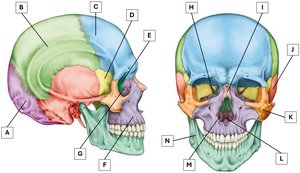
Q2. Name the facial and cranial bones of the skull as labelled in figure 1 below.
Background
Topic: Anatomy of the Skull
This question tests your ability to identify and name the major bones of the human skull, distinguishing between cranial and facial bones.
Key Terms:
Cranial bones: Bones that form the cranium and protect the brain (e.g., frontal, parietal, occipital).
Facial bones: Bones that form the structure of the face (e.g., maxilla, mandible, zygomatic).
Step-by-Step Guidance
Examine the labelled diagram and note the position of each bone. Cranial bones are generally located in the upper and posterior parts of the skull, while facial bones are in the lower and anterior regions.
Recall the names and locations of the cranial bones: frontal, parietal, occipital, temporal, sphenoid, and ethmoid.
Recall the names and locations of the facial bones: maxilla, mandible, zygomatic, nasal, lacrimal, palatine, inferior nasal concha, and vomer.
Match each label (A–N) to the correct bone based on its anatomical location and shape.
Try solving on your own before revealing the answer!
Final Answer:
The labelled bones are: A - Occipital bone, B - Parietal bone, C - Frontal bone, D - Sphenoid bone, E - Ethmoid, F - Maxilla, G - Lacrimal bone, H - Palatine bone, I - Nasal bone, J - Temporal bone, K - Zygomatic bone, L - Bony nasal septum/vomer, M - Inferior nasal concha, N - Mandible.
These labels correspond to the major cranial and facial bones, helping you identify their locations and functions in the skull.
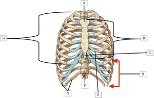
Q6. Name the structures labelled in the diagram below (figure 2).
Background
Topic: Anatomy of the Thoracic Cage
This question tests your ability to identify the main anatomical features of the thoracic cage, including the sternum, ribs, and associated cartilage.
Key Terms:
Sternum: The central bone of the chest, consisting of the manubrium, body, and xiphoid process.
Ribs: Bones forming the sides of the thoracic cage, classified as true, false, and floating ribs.
Costal cartilage: Cartilage connecting ribs to the sternum.
Vertebrae: Bones forming the spine.
Step-by-Step Guidance
Examine the diagram and identify the central bone (sternum) and its parts: manubrium, body, and xiphoid process.
Locate the ribs and distinguish between true, false, and floating ribs based on their attachment to the sternum.
Identify the costal cartilage connecting the ribs to the sternum.
Find the vertebrae at the posterior aspect of the thoracic cage.
Try solving on your own before revealing the answer!
Final Answer:
The labelled structures are: A - Manubrium, B - Sternum, C - Xiphoid process, D - False ribs, E - Costal cartilage, F - Vertebrae, G - Floating ribs, H - True ribs.
These features are essential for understanding the structure and function of the thoracic cage.
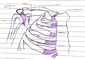
Q8. What bones make up the pectoral girdle? Draw an image and label the main features of these bones.
Background
Topic: Anatomy of the Pectoral Girdle
This question tests your knowledge of the bones forming the pectoral girdle and their anatomical features.
Key Terms:
Pectoral girdle: The set of bones connecting the upper limb to the trunk, including the clavicle and scapula.
Acromioclavicular joint: The joint between the acromion of the scapula and the clavicle.
Humerus: The bone of the upper arm.
Step-by-Step Guidance
Recall that the pectoral girdle consists of the clavicle and scapula.
Identify the acromioclavicular joint, which connects the clavicle and scapula.
Locate the humerus, which articulates with the scapula at the shoulder joint.
Label the main features on your drawing: clavicle, scapula, acromioclavicular joint, and humerus.
Try solving on your own before revealing the answer!
Final Answer:
The bones of the pectoral girdle are the clavicle and scapula. The main features include the acromioclavicular joint, clavicle, scapula, and humerus.
These structures provide support and mobility for the upper limb.
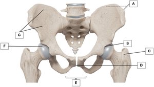
Q10. Identify the following bones of the pelvic girdle by including the correct label from the image below (figure 3).
Background
Topic: Anatomy of the Pelvic Girdle
This question tests your ability to identify and label the bones and features of the pelvic girdle.
Key Terms:
Iliac crest: The upper border of the ilium.
Acetabulum: The socket for the head of the femur.
Ilium: The largest bone of the pelvis.
Femur head: The ball-shaped proximal end of the femur.
Pubic arch: The arch formed by the pubic bones.
Greater trochanter: A large projection on the femur.
Pubic symphysis: The joint between the two pubic bones.
Step-by-Step Guidance
Examine the labelled diagram and locate the iliac crest, acetabulum, ilium, femur head, pubic arch, greater trochanter, and pubic symphysis.
Match each label (A–G) to the correct anatomical feature based on its position and shape.
Recall the function of each feature in supporting the body and facilitating movement.
Try solving on your own before revealing the answer!
Final Answer:
The labelled bones and features are: A - Iliac crest, B - Acetabulum, C - Ilium, D - Pubic symphysis, E - Greater trochanter, F - Pubic arch, G - Femur head.
These features are important for weight-bearing and movement of the lower limb.
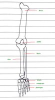
Q11. Draw a leg and label the following bones: Femur; Patella; Fibula; Tibia; Tarsals; Metatarsals; Phalanges.
Background
Topic: Anatomy of the Lower Limb
This question tests your ability to identify and label the bones of the leg and foot.
Key Terms:
Femur: The thigh bone, largest bone in the body.
Patella: The kneecap.
Fibula: The lateral bone of the lower leg.
Tibia: The medial bone of the lower leg.
Tarsals: The bones of the ankle.
Metatarsals: The bones of the foot.
Phalanges: The bones of the toes.
Step-by-Step Guidance
Draw the outline of the leg, including the thigh, knee, lower leg, ankle, and foot.
Label the femur, patella, tibia, and fibula in the correct anatomical positions.
Identify and label the tarsals, metatarsals, and phalanges in the foot.
Count the bones in each section to check your understanding of their arrangement.
Try solving on your own before revealing the answer!
Final Answer:
The labelled bones are: Femur, Patella, Tibia, Fibula, Tarsals, Metatarsals, Phalanges. There are 30 bones in total in the leg.
These bones provide support, movement, and stability for the lower limb.
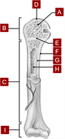
Q12. Name the structures on the diagram (figure 4) of a long bone.
Background
Topic: Anatomy of Long Bones
This question tests your ability to identify the main anatomical features of a long bone, such as the femur or humerus.
Key Terms:
Spongy bone: The porous bone tissue found at the ends of long bones.
Epiphysis: The end part of a long bone.
Diaphysis: The shaft of a long bone.
Articular cartilage: Cartilage covering the ends of bones at joints.
Epiphyseal plate: The growth plate in children and adolescents.
Periosteum: The membrane covering the outer surface of bones.
Compact bone: Dense bone tissue forming the outer layer.
Medullary cavity: The central cavity containing bone marrow.
Distal epiphysis: The end of the bone farthest from the body.
Step-by-Step Guidance
Examine the labelled diagram and identify the spongy bone, epiphysis, diaphysis, articular cartilage, epiphyseal plate, periosteum, compact bone, medullary cavity, and distal epiphysis.
Recall the function of each structure in bone growth, support, and movement.
Match each label (A–I) to the correct anatomical feature based on its position and appearance.
Try solving on your own before revealing the answer!
Final Answer:
The labelled structures are: A - Spongy bone, B - Epiphysis, C - Diaphysis/shaft, D - Articular cartilage, E - Epiphyseal plate, F - Periosteum, G - Compact bone, H - Medullary cavity, I - Distal epiphysis.
These features are essential for understanding bone structure and function.
