 Back
BackStructural Features of Vertebrae: Cervical, Thoracic, and Atlas/Axis
Study Guide - Smart Notes
 Tailored notes based on your materials, expanded with key definitions, examples, and context.
The Skeletal System
Vertebral Structure and Regional Differences
The vertebral column is composed of a series of bones called vertebrae, which are grouped into cervical, thoracic, and lumbar regions. Each region has vertebrae with distinct structural features that reflect their functions and anatomical relationships. The atlas (C1) and axis (C2) are specialized cervical vertebrae that support the skull and allow for head rotation.
General Structure of a Typical Vertebra
Body: The thick, disc-shaped anterior portion that bears weight.
Vertebral Foramen: The opening through which the spinal cord passes.
Pedicle and Lamina: Form the vertebral arch, enclosing the vertebral foramen.
Spinous Process: Posterior projection for muscle and ligament attachment.
Transverse Processes: Lateral projections for muscle and ligament attachment.
Articular Processes (Superior and Inferior): Form joints with adjacent vertebrae.
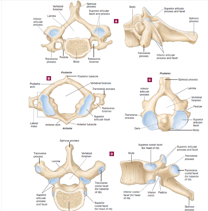
Cervical Vertebrae
Located in the neck region; typically seven in number (C1–C7).
Transverse Foramina: Openings in the transverse processes for vertebral arteries.
Bifid Spinous Process: (Except C7) Split at the tip for muscle attachment.
Atlas (C1): Lacks a body and spinous process; supports the skull and allows nodding motion.
Axis (C2): Has a prominent dens (odontoid process) that acts as a pivot for head rotation.
Atlas (C1) and Axis (C2) Specializations
Atlas (C1): Ring-like structure with anterior and posterior arches, lateral masses, and large superior articular facets for articulation with the occipital condyles of the skull.
Axis (C2): Characterized by the dens, which projects superiorly and articulates with the atlas, allowing rotation of the head ("no" motion).
Thoracic Vertebrae
Located in the upper and mid-back; twelve in number (T1–T12).
Costal Facets: Articulate with the heads and tubercles of ribs.
Spinous Process: Long and points downward.
Body: Heart-shaped and larger than cervical vertebrae.
Key Differences Between Cervical and Thoracic Vertebrae
Feature | Cervical Vertebrae | Thoracic Vertebrae |
|---|---|---|
Body Shape | Small, oval | Larger, heart-shaped |
Spinous Process | Short, often bifid | Long, points downward |
Transverse Foramina | Present | Absent |
Costal Facets | Absent | Present (for ribs) |
Examples and Clinical Relevance
Atlas and Axis: Injury to these vertebrae can impair head movement and spinal cord function.
Thoracic Vertebrae: Fractures may affect rib articulation and respiratory mechanics.
Additional info: The vertebral column provides structural support, protects the spinal cord, and allows flexible movement. Regional differences in vertebrae reflect their roles in supporting the head, facilitating movement, and articulating with ribs.
