 Back
BackStructure and Organization of the Brain
Study Guide - Smart Notes
 Tailored notes based on your materials, expanded with key definitions, examples, and context.
The Brain: Gross Anatomy and Functional Areas
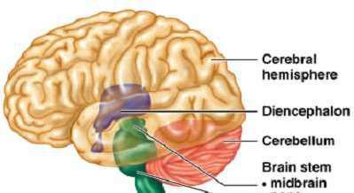
Major Regions of the Brain
The brain is divided into several major regions, each with distinct anatomical and functional characteristics. Understanding these regions is essential for studying the central nervous system.
Cerebral Hemispheres: The largest part of the brain, responsible for higher cognitive functions, voluntary movement, and sensory perception.
Diencephalon: Contains structures such as the thalamus and hypothalamus, which are involved in sensory relay and homeostatic regulation.
Cerebellum: Coordinates voluntary movements and maintains posture and balance.
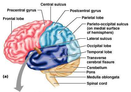
Brain Stem: Includes the midbrain, pons, and medulla oblongata; controls basic life functions such as breathing and heart rate.
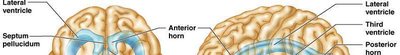
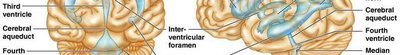
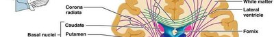
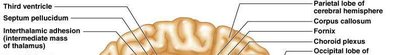
Ventricular System of the Brain
The brain contains a series of interconnected cavities called ventricles, which are filled with cerebrospinal fluid (CSF). These structures help protect the brain and maintain its chemical environment.
Lateral Ventricles: Paired structures located within each cerebral hemisphere.
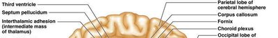
Third Ventricle: Located in the diencephalon, connected to the lateral ventricles by the interventricular foramina.
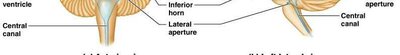
Fourth Ventricle: Located between the brainstem and cerebellum, connected to the third ventricle by the cerebral aqueduct.
Central Canal: Extends from the fourth ventricle into the spinal cord.
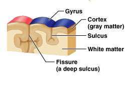
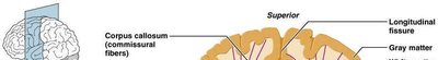
Surface Anatomy of the Cerebrum
The surface of the cerebral hemispheres is highly folded, increasing the surface area for cortical neurons. Key features include gyri, sulci, and fissures.
Gyrus (plural: gyri): Elevated ridges of the cerebral cortex.
Sulcus (plural: sulci): Shallow grooves between gyri.
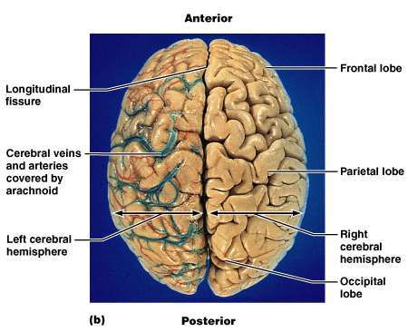
Fissure: Deep grooves that separate major regions of the brain (e.g., longitudinal fissure).
Cortex (gray matter): Outer layer containing neuron cell bodies.
White Matter: Inner region containing myelinated axons.
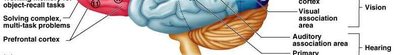
Lobes of the Cerebral Hemispheres
The cerebral hemispheres are divided into lobes, each associated with specific functions.
Frontal Lobe: Involved in voluntary movement, planning, reasoning, and problem-solving.
Parietal Lobe: Processes sensory information such as touch, temperature, and pain.
Temporal Lobe: Responsible for auditory processing and memory.
Occipital Lobe: Primary center for visual processing.
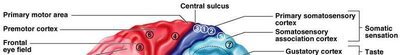
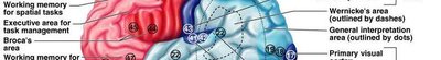
Functional Areas of the Cerebral Cortex
The cerebral cortex contains specialized regions responsible for different sensory and motor functions.
Primary Motor Cortex: Controls voluntary movements.
Premotor Cortex: Plans complex movements.
Primary Somatosensory Cortex: Receives sensory input from the body.
Somatosensory Association Cortex: Integrates sensory information.
Primary Visual Cortex: Processes visual information.
Primary Auditory Cortex: Processes auditory information.
Broca's Area: Involved in speech production.
Wernicke's Area: Involved in language comprehension.
White Matter and Basal Nuclei
White matter consists of myelinated axons that connect different regions of the brain. Basal nuclei are clusters of gray matter deep within the cerebral hemispheres, involved in movement regulation.
Association Fibers: Connect regions within the same hemisphere.
Commissural Fibers: Connect corresponding regions of the two hemispheres (e.g., corpus callosum).
Projection Fibers: Connect the cortex with lower brain regions and the spinal cord.
Basal Nuclei: Include the caudate nucleus, putamen, and globus pallidus; regulate voluntary motor activities.

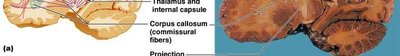
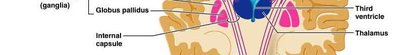
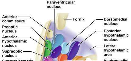
Diencephalon: Thalamus and Hypothalamus
The diencephalon is a central core of brain tissue, including the thalamus and hypothalamus, which are critical for sensory processing and homeostasis.
Thalamus: Acts as a relay station for sensory information traveling to the cerebral cortex.
Hypothalamus: Regulates autonomic functions, endocrine activity, body temperature, hunger, and thirst.
Epithalamus: Contains the pineal gland, which secretes melatonin.


Cerebellum
The cerebellum is located posterior to the brainstem and is responsible for coordinating voluntary movements and maintaining balance and posture.
Anterior and Posterior Lobes: Main functional regions of the cerebellum.
Vermis: Central region connecting the two hemispheres of the cerebellum.
Fissures: Separate the lobes and regions of the cerebellum.
Meninges and Protection of the Brain
The brain is protected by three connective tissue membranes called meninges, as well as the skull and cerebrospinal fluid.
Dura Mater: Tough, outermost layer.
Arachnoid Mater: Middle, web-like layer.
Pia Mater: Thin, innermost layer adhering to the brain surface.
Subarachnoid Space: Contains cerebrospinal fluid and blood vessels.
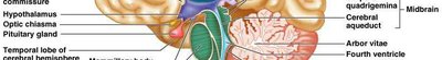
Choroid Plexus and Cerebrospinal Fluid (CSF) Production
The choroid plexus is a network of capillaries and ependymal cells within the ventricles that produces cerebrospinal fluid, which cushions and nourishes the brain.
Ependymal Cells: Line the ventricles and help produce and circulate CSF.
Choroid Plexus: Specialized tissue that filters blood plasma to form CSF.
Summary Table: Major Brain Regions and Functions
Region | Main Function(s) |
|---|---|
Cerebral Hemispheres | Higher cognitive functions, voluntary movement, sensory perception |
Diencephalon | Sensory relay, homeostasis, endocrine regulation |
Cerebellum | Coordination of movement, balance, posture |
Brain Stem | Autonomic functions, relay between brain and spinal cord |
