 Back
BackStudy Guidance for ANP College Course: Anatomical Planes, Body Cavities, and Organ Identification
Study Guide - Smart Notes
 Tailored notes based on your materials, expanded with key definitions, examples, and context.
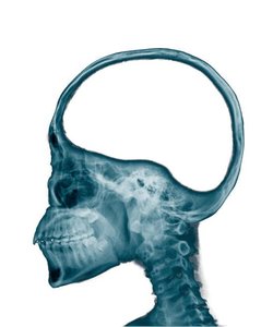
Q1. What type of sections are shown here?
Background
Topic: Anatomical Planes and Sections
This question tests your understanding of anatomical planes used in medical imaging and dissection, such as sagittal, coronal, and transverse sections.
Key Terms:
Sagittal plane: Divides the body into left and right portions.
Coronal (frontal) plane: Divides the body into anterior (front) and posterior (back) portions.
Transverse (horizontal) plane: Divides the body into superior (upper) and inferior (lower) portions.
Step-by-Step Guidance
Observe the orientation of the anatomical structures in the image. Are you seeing a side view, a front/back view, or a top/bottom view?
Identify any visible landmarks (such as the nose, mouth, or spine) to help determine the direction of the section.
Recall the definitions of sagittal, coronal, and transverse planes and match them to the image's perspective.
Consider whether the section divides the body into left/right, front/back, or upper/lower portions.
Try solving on your own before revealing the answer!
Final Answer: Sagittal section
This image shows a sagittal section, which divides the body into left and right portions. The lateral view of the head and neck is characteristic of a sagittal plane.
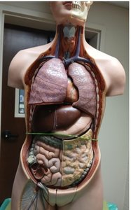
Q2. What three body cavities can you clearly see in these scans?
Background
Topic: Body Cavities
This question tests your knowledge of the major body cavities visible in anatomical models or scans, such as the thoracic, abdominal, and pelvic cavities.
Key Terms:
Thoracic cavity: Contains the heart and lungs.
Abdominal cavity: Contains digestive organs like the stomach, liver, and intestines.
Pelvic cavity: Contains organs such as the urinary bladder and reproductive organs.
Step-by-Step Guidance
Examine the anatomical model for visible organs and their locations.
Identify the boundaries between the thoracic, abdominal, and pelvic cavities.
Recall which organs are found in each cavity to help confirm your identification.
Label each cavity based on the organs and their anatomical positions.
Try solving on your own before revealing the answer!
Final Answer: Thoracic, Abdominal, and Pelvic cavities
The scan clearly shows the thoracic cavity (lungs and heart), abdominal cavity (digestive organs), and pelvic cavity (urinary bladder and reproductive organs).
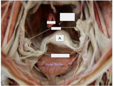
Q3. What is the organ labeled "A" in this cadaver dissection?
Background
Topic: Organ Identification in Dissection
This question tests your ability to identify organs in a cadaver dissection, focusing on anatomical landmarks and relationships.
Key Terms:
Urinary bladder: A hollow organ that stores urine.
Pelvic cavity: The body cavity where the urinary bladder is located.
Step-by-Step Guidance
Locate the label "A" in the image and observe the surrounding structures.
Identify the anatomical region (pelvic cavity) and look for distinguishing features of the organ.
Recall the appearance and function of organs found in the pelvic cavity.
Compare the labeled structure to textbook images or models of pelvic organs.
Try solving on your own before revealing the answer!
Final Answer: Urinary bladder
The organ labeled "A" is the urinary bladder, located in the pelvic cavity and responsible for storing urine.
