 Back
BackStudy Guidance for Chapter 15: The Special Senses (ANP College Course)
Study Guide - Smart Notes
 Tailored notes based on your materials, expanded with key definitions, examples, and context.
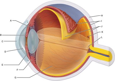
Q4. Assign the letters (A–L) to their corresponding structures of the eye. Refer to Figure 15.4 for help.
Background
Topic: Anatomy of the Eye
This question tests your knowledge of the anatomical structures of the eye and their locations. Understanding these structures is essential for learning about vision and the function of the eye.
Key Terms:
Cornea: Transparent front part of the eye that covers the iris and pupil.
Iris: Colored part of the eye that controls pupil size.
Optic disc: Area where the optic nerve exits the eye; also called the blind spot.
Choroid: Vascular layer between the retina and sclera.
Fovea centralis: Small pit in the retina responsible for sharp central vision.
Ciliary zonule: Fibers that hold the lens in place.
Sclera: White, tough outer layer of the eyeball.
Retina: Light-sensitive layer at the back of the eye.
Ciliary body: Structure containing muscle and processes that control lens shape and produce aqueous humor.
Lens: Transparent structure that focuses light onto the retina.
Anterior segment: Front part of the eye, contains aqueous humor.
Posterior segment: Back part of the eye, contains vitreous humor.
Step-by-Step Guidance
Carefully examine Figure 15.4 and identify each labeled structure (A–L) in the diagram.
Review the definitions and functions of each structure listed above. Match each letter to the correct anatomical term based on its location and appearance in the diagram.
Use the text and figure to confirm your choices. For example, the cornea is the transparent layer at the very front of the eye, while the sclera is the white, outermost layer.
Pay attention to the internal structures, such as the lens, ciliary body, and retina, and how they are positioned relative to each other.
Assign the correct letter to each structure, but do not fill in the final answers yet. Make sure you understand why each structure is located where it is.
Try solving on your own before revealing the answer!
Final Answer:
A: Cornea B: Iris C: Optic disc D: Choroid E: Fovea centralis F: Ciliary zonule G: Sclera H: Retina I: Ciliary body J: Lens K: Anterior segment L: Posterior segment
Each letter corresponds to a specific structure in the eye, as shown in the diagram. Understanding these locations is crucial for learning about vision and eye function.
