 Back
BackStudy Guidance for Pulmonary Volumes, Hemoglobin Saturation, Swallowing, Liver Histology, Small Intestine Structure, and Large Intestine Anatomy
Study Guide - Smart Notes
 Tailored notes based on your materials, expanded with key definitions, examples, and context.
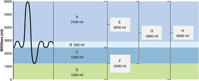
Q1. Identify the pulmonary volumes and capacities labeled A–H in the spirographic record.
Background
Topic: Pulmonary Volumes and Capacities
This question tests your understanding of the different lung volumes and capacities measured during pulmonary function testing. These measurements help assess respiratory health and function.
Key Terms and Formulas:
Tidal Volume (TV): Volume of air inhaled or exhaled with each breath under resting conditions.
Inspiratory Reserve Volume (IRV): Volume of air that can be inspired forcibly beyond the tidal volume.
Expiratory Reserve Volume (ERV): Volume of air that can be expired forcibly beyond the tidal volume.
Residual Volume (RV): Volume of air remaining in the lungs after a forced expiration.
Vital Capacity (VC):
Total Lung Capacity (TLC):
Inspiratory Capacity (IC):
Functional Residual Capacity (FRC):
Step-by-Step Guidance
Examine the spirographic record and locate the labels A–H. Each label corresponds to a specific lung volume or capacity.
Recall the definitions of each volume and capacity. For example, the largest volume (H) is likely the total lung capacity, while the smallest (B) is the tidal volume.
Match the given volumes (e.g., 500 ml, 1200 ml, 3100 ml) to their typical values for a healthy adult to help identify each label.
Use the formulas above to check your assignments. For example, if you know VC, TV, and IRV, you can solve for ERV.
Try solving on your own before revealing the answer!
Final Answer:
A: Inspiratory Reserve Volume (IRV) – 3100 ml
B: Tidal Volume (TV) – 500 ml
C: Expiratory Reserve Volume (ERV) – 1200 ml
D: Residual Volume (RV) – 1200 ml
E: Inspiratory Capacity (IC) – 3600 ml
F: Functional Residual Capacity (FRC) – 2400 ml
G: Vital Capacity (VC) – 4800 ml
H: Total Lung Capacity (TLC) – 6000 ml
These values are based on standard definitions and the typical adult lung volumes. Matching the numbers to the definitions helps you identify each part of the spirogram.
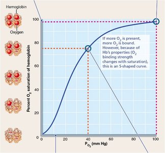
Q2. Explain the significance of the steep portion of the oxygen-hemoglobin dissociation curve at lower partial pressures of O2.
Background
Topic: Oxygen Transport and Hemoglobin Saturation
This question focuses on the oxygen-hemoglobin dissociation curve, which describes how readily hemoglobin binds to and releases oxygen depending on the partial pressure of oxygen ().
Key Terms and Concepts:
Hemoglobin Saturation: The percentage of hemoglobin molecules bound to oxygen.
Partial Pressure of Oxygen (): The pressure exerted by oxygen in a mixture of gases, measured in mm Hg.
Steep Portion of Curve: Indicates that small changes in result in large changes in hemoglobin saturation.
Step-by-Step Guidance
Observe the S-shaped (sigmoidal) curve. The steep portion occurs at lower values (e.g., in tissues).
Understand that in this region, a small drop in leads to a significant release of O2 from hemoglobin.
Relate this to tissue oxygen delivery: as tissues use O2 and drops, hemoglobin releases more O2 efficiently.
Try solving on your own before revealing the answer!
Final Answer:
The steep portion of the curve at lower means that hemoglobin releases oxygen readily where it is needed most (in metabolically active tissues). This ensures efficient oxygen unloading as tissue decreases.
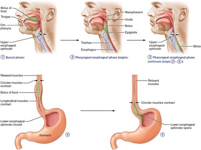
Q3. Match each step of swallowing to its description using the provided diagram.
Background
Topic: Swallowing (Deglutition) Mechanism
This question tests your understanding of the coordinated muscular actions that move food from the mouth to the stomach.
Key Terms:
Bucal Phase: Voluntary phase where the tongue pushes the bolus into the oropharynx.
Pharyngeal-Esophageal Phase: Involuntary phase involving the movement of the bolus through the pharynx and esophagus.
Peristalsis: Wave-like muscle contractions that move the bolus toward the stomach.
Step-by-Step Guidance
Identify each phase in the diagram: buccal (voluntary), pharyngeal-esophageal (involuntary), and the role of sphincters.
Match the muscular actions (e.g., tongue movement, sphincter opening/closing, peristalsis) to the correct step number.
Follow the path of the bolus from the mouth, through the pharynx, into the esophagus, and finally into the stomach.
Try solving on your own before revealing the answer!
Final Answer:
Step 1: Tongue forces bolus into oropharynx (buccal phase)
Step 2: Larynx rises, epiglottis bends to block trachea
Step 3: Upper esophageal sphincter contracts after food enters
Step 4: Peristalsis moves bolus through esophagus
Step 5: Lower esophageal sphincter opens
Each step ensures the safe and efficient movement of food from the mouth to the stomach, preventing aspiration into the airway.
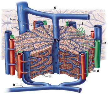
Q4. Label the structures of a liver lobule in the provided diagram.
Background
Topic: Liver Histology
This question focuses on the microscopic anatomy of the liver, specifically the organization of the liver lobule and its associated vessels and cells.
Key Terms:
Central Vein: Drains blood from the lobule.
Portal Triad: Consists of a branch of the hepatic artery, portal vein, and bile duct.
Sinusoids: Capillary-like vessels where blood from the portal triad flows toward the central vein.
Hepatocytes: Liver cells that process blood contents.
Stellate Macrophages (Kupffer Cells): Immune cells in the sinusoids.
Step-by-Step Guidance
Identify the central vein at the center of the lobule.
Locate the portal triads at the corners of the lobule (containing a bile duct, portal vein, and hepatic artery).
Trace the flow of blood from the portal triad through the sinusoids to the central vein.
Identify the hepatocytes and stellate macrophages within the lobule.
Try solving on your own before revealing the answer!
Final Answer:
a: Bile duct
b: Portal vein
c: Hepatocyte
d: Portal triad
e: Sinusoid
f: Bile canaliculus
g: Central vein
h: Stellate macrophage (Kupffer cell)
Understanding the structure of the liver lobule is essential for grasping how the liver processes blood and produces bile.
Q5. Assign the structural modifications of the small intestine wall (A–C) to their correct terms.
Background
Topic: Small Intestine Structure and Function
This question tests your knowledge of the adaptations that increase the surface area for absorption in the small intestine.
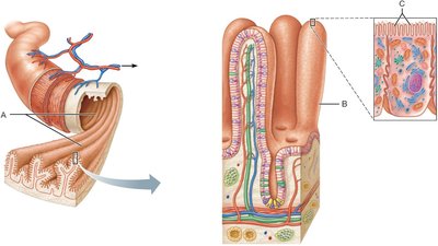
Key Terms:
Circular Folds (A): Deep folds of the mucosa and submucosa that slow the movement of chyme and increase absorption time.
Villi (B): Fingerlike projections of the mucosa that increase surface area for absorption.
Microvilli (C): Tiny projections on the surface of enterocytes that form the brush border and further increase surface area.
Step-by-Step Guidance
Examine the diagram and identify the three structural modifications labeled A, B, and C.
Recall the function of each structure in increasing the absorptive surface area of the small intestine.
Match each label to its correct term based on its appearance and location in the diagram.
Try solving on your own before revealing the answer!
Final Answer:
A: Circular folds
B: Villi
C: Microvilli
These modifications work together to maximize nutrient absorption in the small intestine.
Q6. Label the major regions of the large intestine in the provided diagram.
Background
Topic: Large Intestine Anatomy
This question focuses on identifying the main anatomical regions of the large intestine, which is important for understanding its role in water absorption and feces formation.
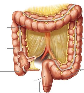
Key Terms:
Cecum: The first part of the large intestine.
Ascending Colon: Travels up the right side of the abdominal cavity.
Transverse Colon: Runs across the abdominal cavity.
Descending Colon: Travels down the left side of the abdominal cavity.
Sigmoid Colon: S-shaped region leading to the rectum.
Rectum: The final straight portion of the large intestine.
Anal Canal: The terminal part of the large intestine.
Step-by-Step Guidance
Identify each labeled region in the diagram based on its position and shape.
Recall the order in which material passes through the large intestine: cecum → ascending colon → transverse colon → descending colon → sigmoid colon → rectum → anal canal.
Match each label to the correct anatomical term.
Try solving on your own before revealing the answer!
Final Answer:
a: Cecum
b: Ascending colon
c: Transverse colon
d: Descending colon
e: Sigmoid colon
f: Rectum
g: Anal canal
Knowing these regions helps you understand the flow of material and the functions of the large intestine.
