 Back
CNS part 1 & 2
Study Guide - Smart Notes
 Tailored notes based on your materials, expanded with key definitions, examples, and context.
The Central Nervous System: Overview
Definition and Components
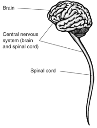
The central nervous system (CNS) is the primary control center of the body, consisting of the brain and spinal cord. It is responsible for processing sensory information, generating thoughts, emotions, and controlling motor functions.
Brain: The organ responsible for cognition, emotion, and voluntary actions.
Spinal cord: The pathway for transmitting information between the brain and the rest of the body.
Directional Terminology in CNS Anatomy
Rostral and Caudal
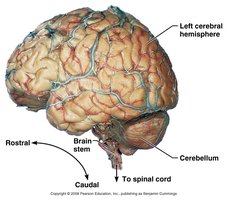
Directional terms are used to describe locations within the CNS:
Rostral: Toward the nose or forehead (synonymous with anterior).
Caudal: Toward the tail or spinal cord (synonymous with posterior).
Gray and White Matter in the CNS
Structural Organization
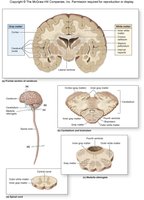
The CNS is organized into two main tissue types:
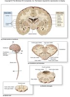
Gray matter: Composed of neuronal cell bodies, dendrites, and unmyelinated axons. In the brain, the outer layer is called the cortex, and deep regions are called nuclei.
White matter: Consists mainly of myelinated axons, responsible for rapid communication between different brain regions.
Major White Matter Structures
Corpus callosum: Connects the left and right cerebral hemispheres.
Internal capsule: Connects the brainstem and cerebral cortex.
Septum pellucidum: Membrane connecting corpus callosum and fornix.
Basic Organization of the Brain
Major Regions
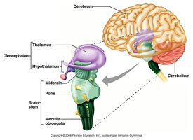
The brain is divided into four main regions:
Brain Stem
Cerebellum
Diencephalon
Cerebrum
Cerebrum
Structure and Surface Features
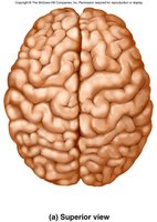
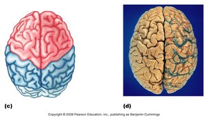
The cerebrum is the largest and most rostral part of the brain, covering the diencephalon and brainstem. It is divided into two hemispheres and makes up about 80% of total brain mass.
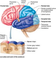
Gyri: Elevated ridges on the brain surface.
Sulci: Shallow grooves adjacent to gyri.
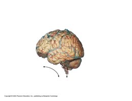
Fissures: Deep grooves separating major brain regions.
Major Fissures
Transverse cerebral fissure: Separates cerebral hemispheres from the cerebellum.
Longitudinal fissure: Separates right and left hemispheres.
Organization of the Cerebral Hemispheres
Outer gray matter (cerebral cortex)
White matter internal to the cortex
Gray matter within the white matter (deep nuclei)
Cerebral Cortex
Structure and Function
The cerebral cortex is a thin layer (2-4 mm) of gray matter responsible for consciousness, motor initiation, communication, memory, and comprehension.
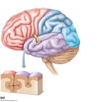
Lobes of the Cerebral Cortex
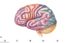
The cortex is divided into five lobes, named after the overlying skull bones:
Frontal lobe
Parietal lobe
Temporal lobe
Occipital lobe
Insula (hidden beneath other lobes)
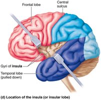
Functional Areas of the Cerebral Cortex
Sensory areas: Allow conscious perception of sensations.
Motor areas: Control voluntary body movements.
Association areas: Perform higher processing and integration of information.
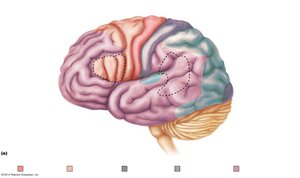
Sensory Areas and Their Functions
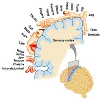
Parietal lobe: Primary somatosensory cortex (touch, pressure, temperature).
Occipital lobe: Primary visual cortex (visual stimuli).
Temporal lobe: Primary auditory cortex (auditory stimuli).
Insula: Gustatory cortex (taste) and sense of balance.
Motor Areas
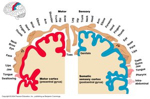
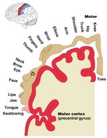
Primary motor cortex: Located on the precentral gyrus; initiates voluntary movement.
Premotor cortex: Coordinates complex movements and relays information to the primary motor cortex.
Homunculus: Spatial Representation
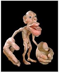
The motor and sensory homunculus are maps on the cortex showing the representation of body parts. Larger areas indicate greater sensitivity or motor control.
Somatosensory Association Area
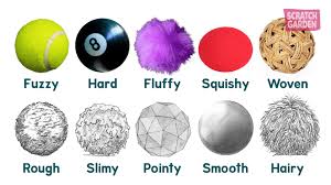
Located posterior to the primary somatosensory cortex, this area integrates sensory information for object recognition, including size, texture, and relationship of felt objects.
Broca's and Wernicke's Areas
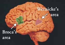
Broca's area: Motor production of speech; damage results in inability to produce speech.
Wernicke's area: Language comprehension; damage results in loss of comprehension but intact speech production.
Visual and Auditory Areas
Primary visual cortex: Receives visual information from the retinas.
Visual association area: Interprets visual stimuli using past experiences.
Primary auditory cortex: Interprets information from the inner ear.
Auditory association area: Stores memories of sound and music.
Other Sensory Areas
Vestibular cortex: Conscious awareness of balance.
Olfactory cortex: Awareness of odors.
Gustatory cortex: Perception of taste.
Visceral sensory area: Perception of visceral sensations.
Multimodal Association Areas
Anterior association area (prefrontal cortex): Intelligence, cognition, personality, planning.
Posterior association area: Pattern and face recognition, spatial localization, language understanding.
Limbic association area: Emotional impact for memories and experiences.
Cerebral White Matter
Structure and Function
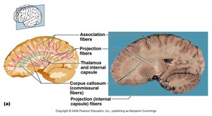
White matter lies deep to the cortex and is composed of myelinated axons. It enables rapid communication between brain regions. The corpus callosum is a major tract connecting the two hemispheres.
Diencephalon
Components and Functions
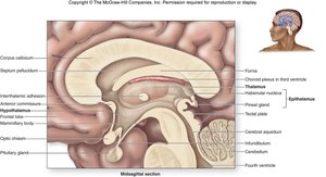
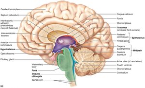
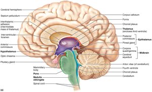
The diencephalon forms the central core of the forebrain and consists of:
Thalamus: Major relay for sensory information (except olfaction).
Hypothalamus: Controls autonomic functions, endocrine system, body temperature, hunger, thirst, and motivational behaviors.
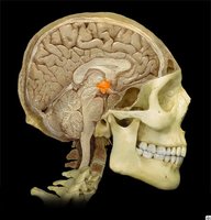
Epithalamus: Contains the pineal gland, which secretes melatonin for sleep regulation.
Brain Stem
Structure and Divisions
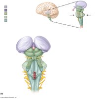
The brain stem is the most caudal region of the brain and consists of:
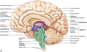
Midbrain: Contains cerebral peduncles, superior and inferior colliculi (visual and auditory reflexes).
Pons: Connects brainstem and cerebellum; involved in movement coordination and respiration.
Medulla oblongata: Contains autonomic nuclei for vital functions (respiration, cardiac function, blood pressure).
Cerebellum
Structure and Function
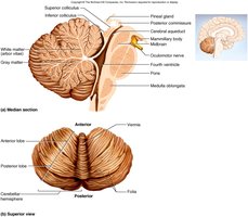
The cerebellum is located dorsal to the pons and medulla. It has two hemispheres connected by the vermis and coordinates body movements, posture, and balance. It also stores procedural (motor) memory.
Reticular Formation
Reticular Activating System (RAS)
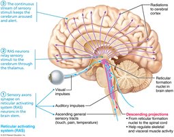
The reticular formation is a network of neurons in the brainstem that maintains consciousness and alertness. The RAS filters sensory input and is inhibited by sleep, alcohol, and drugs. Severe injury can result in coma.
Higher Mental Functions
Memory
Memory involves storage and retrieval of information. Types include:
Declarative memory: Facts, names, dates.
Procedural memory: Skills (e.g., playing piano).
Motor memory: Motor skills (e.g., riding a bike).
Emotional memory: Experiences linked to emotions.
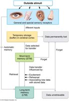
Memory consolidation is the process of converting short-term memory to long-term memory, involving the hippocampus, thalamus, and prefrontal cortex.
Sleep and Sleep-Wake Cycles
Types and Importance of Sleep
Sleep is regulated by circadian rhythms and the RAS. Types include:
Non-rapid eye movement (NREM): Four stages, from light to deep sleep.
Rapid eye movement (REM): Dreaming, increased visceral activity, skeletal muscle inhibition.
REM sleep is important for memory consolidation and emotional processing. Sleep requirements decline with age.
Electroencephalogram (EEG)
Recording Brain Activity
An EEG records electrical activity in the brain and is used to diagnose cerebral dysfunction, brain death, and sleep patterns.
