 Back
BackStudy Guide: Anatomy and Physiology of the Heart (Cardiovascular System)
Study Guide - Smart Notes
 Tailored notes based on your materials, expanded with key definitions, examples, and context.
Heart Anatomy and Cardiovascular System
Overview of the Cardiovascular System
The cardiovascular system is responsible for transporting blood, nutrients, gases, and wastes throughout the body. It consists of three main components: blood, blood vessels (arteries, veins, capillaries), and the heart.
Blood: Carries oxygen, nutrients, and waste products.
Blood Vessels: Arteries carry blood away from the heart, veins return blood to the heart, and capillaries allow exchange between blood and tissues.
Heart: The muscular organ that pumps blood through the circulatory system.
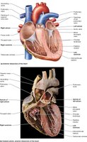
Location and Structure of the Heart
The heart is located in the mediastinum, within the pericardial cavity. Its apex points downward and to the left, while the base is positioned superiorly. The heart is roughly the size of a fist.
Mediastinum: Central compartment of the thoracic cavity.
Pericardial cavity: Space surrounding the heart, filled with pericardial fluid.
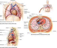
Pericardium and Layers of the Heart Wall
The heart is enclosed by the pericardium, which consists of two main layers: the fibrous pericardium and the serous pericardium. The serous pericardium is further divided into parietal and visceral layers, with the pericardial cavity between them.
Fibrous pericardium: Tough, outer layer that protects and anchors the heart.
Serous pericardium: Double-layered membrane; parietal layer lines the cavity, visceral layer (epicardium) covers the heart.
Pericardial fluid: Reduces friction during heart movements.

Layers of the Heart Wall
The heart wall is composed of three layers, each with distinct functions:
Epicardium (visceral pericardium): Outer layer, provides protection.
Myocardium: Middle, muscular layer responsible for contraction.
Endocardium: Inner layer, lines chambers and valves.

Heart Circuits and Blood Flow
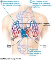
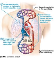
Pulmonary and Systemic Circuits
The heart pumps blood through two main circuits: the pulmonary circuit and the systemic circuit.
Pulmonary circuit: Carries deoxygenated blood from the right side of the heart to the lungs and returns oxygenated blood to the left side.
Systemic circuit: Distributes oxygenated blood from the left side of the heart to the body and returns deoxygenated blood to the right side.
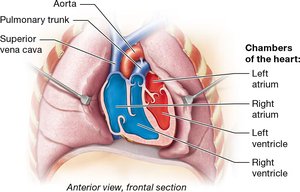
Basic Heart Anatomy and Blood Pathway
The heart has four chambers: right atrium, right ventricle, left atrium, and left ventricle. Blood flows through these chambers in a specific sequence, passing through valves that prevent backflow.
Right Atrium: Receives deoxygenated blood from the body via the superior/inferior vena cava.
Right Ventricle: Pumps blood to the lungs via the pulmonary trunk and arteries.
Left Atrium: Receives oxygenated blood from the lungs via pulmonary veins.
Left Ventricle: Pumps blood to the body via the aorta.
Heart Valves
Valves ensure unidirectional blood flow and prevent backflow:
Atrioventricular (AV) valves: Located between atria and ventricles (tricuspid on right, bicuspid/mitral on left).
Semilunar valves: Located between ventricles and major arteries (pulmonary and aortic valves).
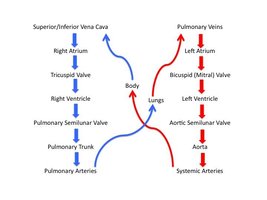
Blood Flow Through the Heart
Blood follows a specific pathway through the heart, passing through chambers and valves:
Body → Superior/Inferior Vena Cava → Right Atrium → Tricuspid Valve → Right Ventricle → Pulmonary Semilunar Valve → Pulmonary Trunk → Pulmonary Arteries → Lungs
Lungs → Pulmonary Veins → Left Atrium → Bicuspid (Mitral) Valve → Left Ventricle → Aortic Semilunar Valve → Aorta → Body
Heart Disorders
Valvular Heart Diseases
Valvular diseases impair the function of heart valves, leading to insufficient closure or stenosis (narrowing due to calcium deposits).
Insufficiency: Valve cannot close fully, causing regurgitation.
Stenosis: Valve opening is narrowed, restricting blood flow.
Symptoms: May include fatigue, shortness of breath, and heart murmurs.
Treatments: Range from medication to surgical repair or replacement.
Coronary Artery Disease
Coronary artery disease is caused by plaque buildup in coronary arteries, leading to reduced blood flow (myocardial ischemia) and potentially myocardial infarction (heart attack).
Risk factors: Include high cholesterol, hypertension, smoking, diabetes, and family history.
Symptoms: Chest pain, shortness of breath, fatigue.
Treatments: Lifestyle changes, medications, angioplasty, or bypass surgery.
Electrical Physiology of the Heart
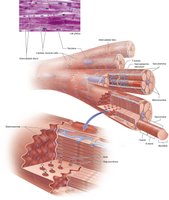
Cardiac Muscle Histology
Cardiac muscle cells are single-nucleated, branched, striated, and connected by intercalated discs, forming a functional syncytium.
Intercalated discs: Specialized junctions for rapid electrical communication.
Functional syncytium: Allows coordinated contraction of the heart.
Cardiac Muscle Cells: Pacemaker and Contractile Cells
Cardiac muscle is intrinsic and autorhythmic, meaning it can generate action potentials without nervous system stimulation.
Pacemaker cells: 1% of cardiac cells, spontaneously generate action potentials.
Contractile cells: 99% of cardiac cells, receive action potentials and contract.
Action Potentials in Cardiac Cells
Cardiac action potentials involve depolarization, repolarization, and hyperpolarization phases, regulated by ion channels.
Depolarization: Na+ channels open, membrane potential rises from -70 to +30 mV.
Repolarization: K+ channels open, membrane potential falls from +30 to -90 mV.
Hyperpolarization: Na+/K+ exchange pump resets membrane potential.
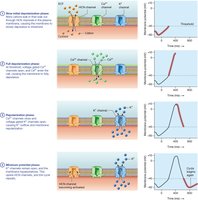
Action Potential in Pacemaker Cells
Pacemaker cells have unique action potential phases:
Initial depolarization: HCN channels allow Na+ influx, less K+ efflux (-60 to -40 mV).
Full depolarization: Ca2+ channels open, Ca2+ enters (-40 to +10 mV).
Repolarization: Ca2+ channels close, K+ channels open (+10 to -40 mV).
Hyperpolarization: K+ channels remain open until -60 mV.
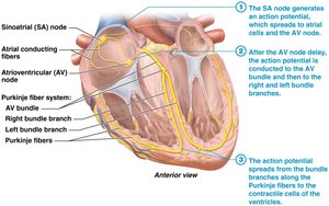
Cardiac Conduction System
The cardiac conduction system consists of interconnected pacemaker cells that coordinate heartbeats.
Sinoatrial (SA) node: Primary pacemaker (~70 BPM).
Atrioventricular (AV) node: Delays impulse.
AV bundle, bundle branches, Purkinje fibers: Distribute impulse to ventricles.
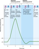
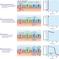
Action Potential in Contractile Cells
Contractile cells have a distinct action potential with a plateau phase:
Rapid depolarization: Na+ channels open (-85 to +20 mV).
Initial repolarization: Na+ channels close, some K+ channels open (+20 to 0 mV).
Plateau: Ca2+ channels open, K+ leaves (sustained at 0 mV).
Repolarization: Na+ and Ca2+ channels close, more K+ channels open (0 to -85 mV).
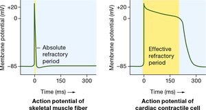
Plateau Phase and Refractory Period
The plateau phase prolongs the action potential, preventing tetanus and allowing proper filling and ejection of blood.
Skeletal muscle AP: 2-5 ms
Cardiac AP: 200-300 ms
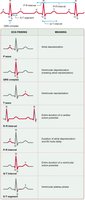
Electrocardiogram (ECG)
An ECG measures the electrical activity of the heart, showing depolarization and repolarization waves.
P Wave: Atrial depolarization
QRS Complex: Ventricular depolarization
T Wave: Ventricular repolarization
ECG Intervals and Segments
Intervals include at least one wave, while segments do not. Key intervals and segments:
R-R interval: Determines heart rate
P-R interval: Time from atrial to ventricular depolarization
Q-T interval: Duration of ventricular activity
S-T segment: Time between ventricular depolarization and repolarization

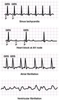
Dysrhythmias
Dysrhythmias are disturbances in heart rate or conduction pathways.
Bradycardia: Heart rate < 60 BPM
Tachycardia: Heart rate > 100 BPM
Heart block: Impaired conduction
Fibrillation: Uncoordinated contractions (atrial or ventricular)
Mechanical Physiology of the Heart
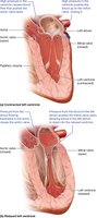
Pressure Changes and Blood Movement
Blood moves through the heart due to pressure gradients created by contraction (systole) and relaxation (diastole). Valves open and close in response to these changes.
Systole: Contraction phase, increases pressure, blood ejected
Diastole: Relaxation phase, decreases pressure, chambers fill
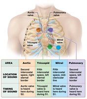
Heart Sounds
Heart sounds are produced by vibrations as valves close:
S1 "lubb": Closure of AV valves
S2 "dupp": Closure of semilunar valves
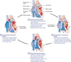
Cardiac Cycle
The cardiac cycle describes events from one heartbeat to the next, divided into four phases:
Ventricular filling: Blood flows into ventricles
Isovolumetric contraction: Ventricles contract, all valves closed
Ventricular ejection: Blood pumped out
Isovolumetric relaxation: Ventricles relax, all valves closed
Cardiovascular Regulation
Neural and Endocrine Regulation
Heart rate and contractility are regulated by the nervous and endocrine systems:
Sympathetic nervous system: Norepinephrine increases heart rate and contractility
Parasympathetic nervous system: Acetylcholine decreases heart rate and contractility
Endocrine system: Hormones (Epi/NE, T3/T4, glucagon) increase heart rate and contractility, effects last longer
Other factors: Body temperature, age, physical fitness
Summary Table: Heart Anatomy and Function
Structure | Function |
|---|---|
Right Atrium | Receives deoxygenated blood from body |
Right Ventricle | Pumps blood to lungs |
Left Atrium | Receives oxygenated blood from lungs |
Left Ventricle | Pumps blood to body |
AV Valves | Prevent backflow into atria |
Semilunar Valves | Prevent backflow into ventricles |
Key Equations
Cardiac Output:
Stroke Volume:
Additional info:
Some details about symptoms and treatments of disorders were inferred based on standard academic knowledge.
Table entries and equations were added for completeness and clarity.
