 Back
BackStudy Guide: Anatomy & Physiology of the Respiratory System
Study Guide - Smart Notes
 Tailored notes based on your materials, expanded with key definitions, examples, and context.
Respiratory System Overview
Functions of the Respiratory System
The respiratory system is essential for gas exchange, protection, and communication. It provides oxygen to the body and removes carbon dioxide, while also protecting respiratory surfaces and facilitating speech and smell.
Gas Exchange: Transfers oxygen from air to blood and removes carbon dioxide.
Air Movement: Moves air into and out of alveoli.
Protection: Protects surfaces from dehydration, temperature changes, and pathogens.
Speech: Vocal cords in the larynx produce sound.
Smell: Olfactory receptors in the nasal cavity aid in the sense of smell.
Cellular Respiration
Cellular respiration is the process by which cells consume oxygen and produce carbon dioxide and ATP, the energy currency of the cell.
Oxygen Consumption: Cells use O2 to generate ATP.
Carbon Dioxide Production: CO2 is a waste product of cellular metabolism.
Anatomy of the Respiratory System
Organs of the Respiratory System
The respiratory system consists of several organs that work together to facilitate breathing and gas exchange.
Nose: Entry point for air; moistens, warms, and filters air.
Nasal Cavity & Paranasal Sinuses: Air passageways; produce mucus and lighten skull.
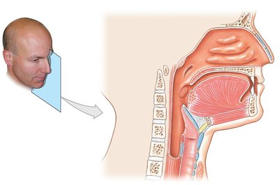
Pharynx: Connects nasal cavity to oral cavity; divided into nasopharynx, oropharynx, and laryngopharynx.
Larynx: Contains vocal cords; prevents food from entering the airway.
Trachea: Windpipe; held open by C-shaped cartilages.
Bronchi and Bronchioles: Branching airways leading to lungs.
Lungs and Alveoli: Main site of gas exchange.
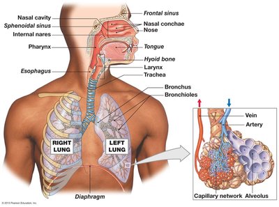
Functional Organization: Conducting vs Respiratory Zone
The respiratory system is divided into two functional zones: the conducting zone and the respiratory zone.
Conducting Zone: Includes nose, nasal cavity, pharynx, trachea, bronchi, and bronchioles. Conducts air to the respiratory zone.
Respiratory Zone: Includes respiratory bronchioles, alveolar ducts, alveolar sacs, and alveoli. Site of gas exchange.
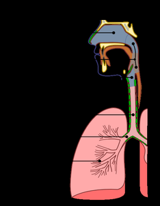
Structural Organization: Upper and Lower Respiratory Tracts
The respiratory tract is structurally divided into upper and lower regions.
Upper Respiratory Tract: Nose, nasal cavity, pharynx, larynx.
Lower Respiratory Tract: Trachea, bronchi, lungs.
Respiratory Mucosa
The conducting zone is lined with respiratory mucosa, which consists of pseudostratified ciliated columnar epithelium and goblet cells.
Function: Traps and removes particles, moistens and warms air.
Nose and Nasal Cavity
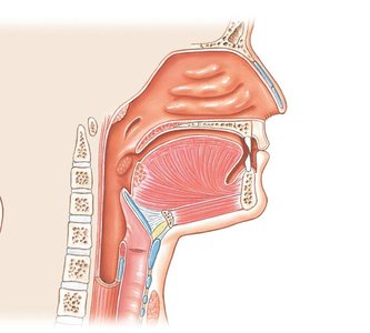
The nose and nasal cavity filter, moisten, and warm incoming air. The nasal conchae create turbulence, increasing contact with mucosa.
Structure: External nares, nasal septum, internal nares, palates, nasal conchae.
Function: Resonating chamber for speech, houses olfactory receptors.
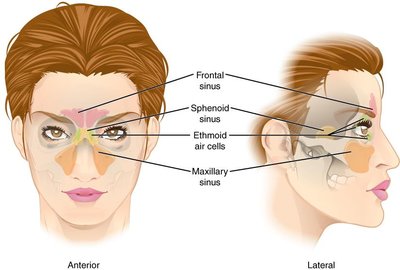
Paranasal Sinuses
Paranasal sinuses are air-filled cavities that produce mucus and lighten the skull.
Types: Frontal, sphenoid, ethmoid, maxillary sinuses.
Pharynx
The pharynx is a funnel-shaped passageway connecting the nasal cavity to the oral cavity. It is divided into three sections: nasopharynx, oropharynx, and laryngopharynx.
Larynx
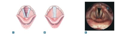
The larynx is composed of several cartilages and houses the vocal cords. It prevents food from entering the airway and produces sound.
Unpaired Cartilages: Epiglottis, thyroid cartilage, cricoid cartilage.
Paired Cartilages: Arytenoid, corniculate, cuneiform cartilages.
Vocal Cords: True vocal cords (sound production), false vocal cords (prevent foreign objects).
Trachea and Bronchi
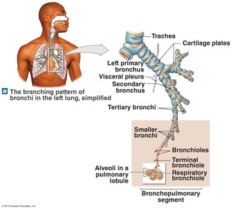
The trachea is supported by C-shaped cartilages and leads to the bronchi, which branch into smaller airways.
Primary Bronchi: Right and left main bronchi.
Secondary Bronchi: Enter lung lobes.
Tertiary Bronchi: Further branching.
Bronchioles: Smallest airways; terminal bronchioles mark the end of the conducting zone.
Alveoli and Respiratory Membrane
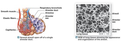
Alveoli are the primary site of gas exchange. The respiratory membrane consists of alveolar and capillary walls plus basement membranes, allowing diffusion of gases.
Cell Types: Type I (gas exchange), Type II (reduce surface tension), alveolar macrophages (remove particles).
Elastic Fibers: Allow expansion and recoil.
Gross Anatomy of the Lungs
Lobes and Pleura
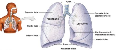
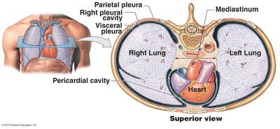
The lungs are divided into lobes and covered by pleura. The right lung has three lobes; the left lung has two lobes and a cardiac notch.
Parietal Pleura: Lines thoracic cavity wall.
Visceral Pleura: Covers lungs.
Pleural Cavity: Space between pleurae.
Pulmonary Ventilation
Mechanics of Breathing
Pulmonary ventilation relies on pressure gradients created by changes in thoracic volume.
Inhalation: Contraction of diaphragm and external intercostals lowers air pressure in lungs, drawing air in.
Exhalation: Relaxation increases pressure, expelling air.
Ventilation Equation
Pulmonary ventilation is calculated as:
Respiratory Rate: Breaths per minute.
Tidal Volume: Volume of air per breath.
Equation:
Gas Exchange and Transport
Partial Pressure and Gas Exchange
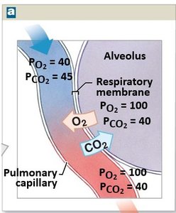
Gas exchange is driven by partial pressure differences of oxygen and carbon dioxide between alveoli and capillaries.
Partial Pressure: Pressure exerted by each gas in a mixture.
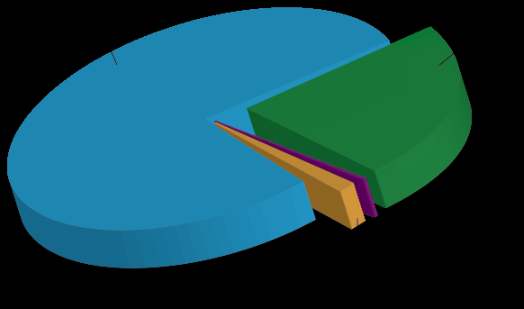
Atmospheric Example: 79% nitrogen, 21% oxygen, <1% carbon dioxide.
External and Internal Respiration
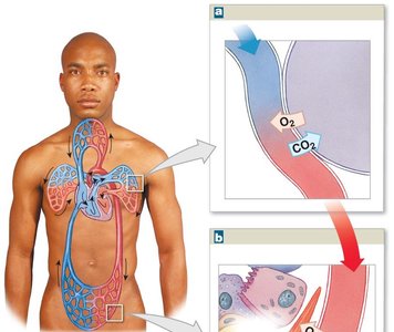
External respiration occurs in the lungs; internal respiration occurs in peripheral tissues.
External Respiration: O2 moves from alveoli to blood; CO2 moves from blood to alveoli.
Internal Respiration: O2 moves from blood to tissues; CO2 moves from tissues to blood.
Gas Transport: Oxygen and Carbon Dioxide
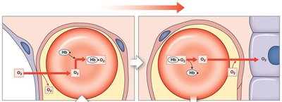
Oxygen is transported mainly by hemoglobin; carbon dioxide is transported in three ways.
Oxygen: 1% dissolved in plasma, 99% bound to hemoglobin.
Carbon Dioxide: 7% dissolved in plasma, 23% bound to hemoglobin, 70% as bicarbonate ions.
Carbon Dioxide Transport Equation
Carbon dioxide is converted to bicarbonate in red blood cells by carbonic anhydrase:

Summary Table: Respiratory System Zones
Zone | Structures | Function |
|---|---|---|
Conducting Zone | Nose, nasal cavity, pharynx, trachea, bronchi, bronchioles | Conducts air to respiratory zone |
Respiratory Zone | Respiratory bronchioles, alveolar ducts, alveolar sacs, alveoli | Gas exchange |
Summary Table: Gas Transport Mechanisms
Gas | Transport Method | Percentage |
|---|---|---|
Oxygen | Dissolved in plasma | ~1% |
Oxygen | Bound to hemoglobin | ~99% |
Carbon Dioxide | Dissolved in plasma | 7% |
Carbon Dioxide | Bound to hemoglobin | 23% |
Carbon Dioxide | As bicarbonate ions | 70% |
Additional info: Academic context was added to clarify the functions, anatomy, and physiology of the respiratory system, as well as to expand on gas exchange and transport mechanisms.
