 Back
BackStudy Guide: Anatomy and Physiology of the Eye (Special Senses)
Study Guide - Smart Notes
 Tailored notes based on your materials, expanded with key definitions, examples, and context.
The Special Senses: The Eye
Surface Anatomy of the Eye
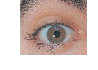
The human eye is a complex organ responsible for vision, and its surface anatomy includes several protective and functional structures.
Eyelids: Protect the eye anteriorly and help spread tears.
Eyebrows: Shade the eye and prevent perspiration from reaching the eye.
Eyelashes: Contain nerve endings that initiate reflex blinking.
Sclera: The white part of the eye, covered by conjunctiva.
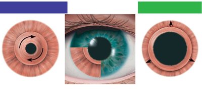
Iris: The colored part of the eye, controls pupil size.
Pupil: The central opening that regulates the amount of light entering the eye.
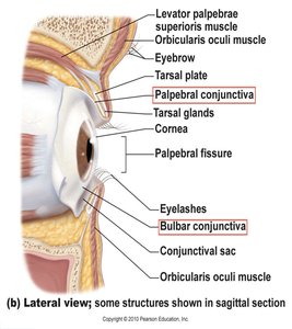
Conjunctiva
The conjunctiva is a transparent membrane that lines the eyelids and covers the white of the eye, producing a lubricating mucous secretion.
Palpebral conjunctiva: Lines the eyelids.
Bulbar conjunctiva: Covers the white of the eyes.
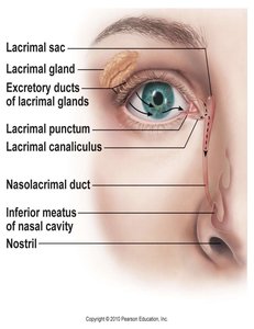
Lacrimal Apparatus
The lacrimal apparatus produces and drains tears, which lubricate and protect the eye.
Lacrimal gland: Produces lacrimal secretion (tears).
Lacrimal puncta: Openings that collect tears.
Lacrimal canaliculi: Channels that drain tears into the lacrimal sac.
Nasolacrimal duct: Drains tears into the nasal cavity.
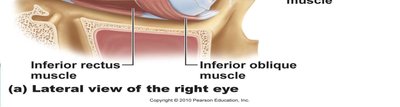
Extrinsic Eye Muscles
Muscles and Their Actions
Six extrinsic muscles control the movement of the eyeball, allowing precise tracking and positioning.
Superior rectus: Elevates the eye.
Inferior rectus: Depresses the eye.
Lateral rectus: Moves the eye laterally.
Medial rectus: Moves the eye medially.
Superior oblique: Depresses the eye and turns it laterally.
Inferior oblique: Elevates the eye and turns it laterally.
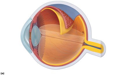
Structure of the Eyeball
Layers of the Eyeball
The wall of the eyeball consists of three layers: fibrous, vascular, and inner (retina). The internal cavity is filled with fluids called humors, and the lens separates the internal cavity into anterior and posterior segments.
Fibrous layer: Sclera and cornea.
Vascular layer (Uvea): Choroid, ciliary body, and iris.
Inner layer: Retina.

Fibrous Layer
The fibrous layer provides protection and shape to the eyeball.
Sclera: Protects and shapes the eyeball; anchors extrinsic eye muscles.
Cornea: Bends light as it enters the eye; contains pain receptors.

Vascular Layer (Uvea)
The vascular layer supplies blood and controls the shape of the lens and pupil.
Choroid: Supplies blood to all layers; absorbs light.
Ciliary body: Controls lens shape; secretes fluid; holds lens in position.
Iris: Colored part; regulates pupil size.


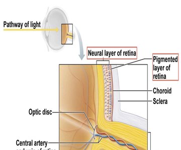
Sensory Layer: Retina
Structure and Function
The retina is a delicate two-layered membrane responsible for photoreception and signal processing.
Pigmented layer: Absorbs light, stores vitamin A.
Neural layer: Contains photoreceptors (rods and cones), bipolar cells, ganglion cells, amacrine cells, and horizontal cells.

Photoreceptors
Photoreceptors are specialized cells that transduce light energy into neural signals.
Rods: More numerous in the peripheral retina; operate in dim light; provide non-color vision.
Cones: Concentrated in the fovea centralis; operate in bright light; provide high-acuity color vision.
Blood Supply to the Retina
The retina receives blood from two sources: the choroid (outer third) and the central artery and vein of the retina (inner two-thirds).
Choroid: Supplies photoreceptors.
Central artery and vein: Supply inner retina.

Internal Chambers and Fluids
Vitreous and Aqueous Humor
The eye contains two main fluids: vitreous humor in the posterior segment and aqueous humor in the anterior segment.
Vitreous humor: Maintains eye shape and optical properties.
Aqueous humor: Supplies nutrients and oxygen to lens and cornea; drains via scleral venous sinus.
Glaucoma: Caused by blocked drainage of aqueous humor, leading to compression of retina and optic nerve.

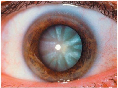
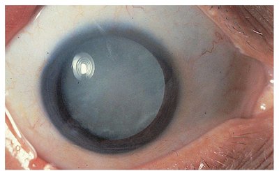
Lens
Structure and Function
The lens is a biconvex, transparent, flexible, and avascular structure that focuses light on the retina. It becomes denser and less elastic with age, leading to conditions such as cataracts.
Cataracts: Clouding of the lens, often age-related or due to diabetes, smoking, or sunlight exposure.
Light and Vision
Properties of Light
Vision depends on the eye's response to visible light, which is a small portion of the electromagnetic spectrum. Light is composed of photons that travel in a wavelike fashion.
Refraction: Bending of light rays as they pass through different media.
Convex lens: Bends light so rays converge at a focal point; image formed is inverted.
Pathway of Light and Focusing
Light enters the eye through the cornea, aqueous humor, lens, and vitreous humor before reaching the retina. Focusing for distant and close vision involves changes in lens shape and pupil size.
Distant vision: Lens is flattened; ciliary muscles relaxed.
Close vision: Lens bulges; ciliary muscles contract.
Accommodation: Changing lens shape for near vision.
Presbyopia: Loss of accommodation with age.
Convergence: Medial rotation of eyeballs for near objects.
Common Vision Disorders
Myopia (nearsightedness): Focal point in front of retina; corrected with concave lens.
Hyperopia (farsightedness): Focal point behind retina; corrected with convex lens.
Astigmatism: Unequal curvatures; corrected with cylindrical lenses or laser procedures.
Phototransduction and Visual Pathways
Phototransduction
Phototransduction is the process by which photoreceptors convert light into electrical signals.
Rhodopsin: Visual pigment in rods; consists of retinal and opsin.
Pigment synthesis: Rhodopsin forms in the dark.
Pigment bleaching: Light absorption causes rhodopsin breakdown.
Pigment regeneration: Rhodopsin is reformed after bleaching.
Visual Pathways
Axons of retinal ganglion cells form the optic nerve, which transmits visual information to the brain. Medial fibers cross at the optic chiasma, and most fibers continue to the lateral geniculate body of the thalamus, then to the primary visual cortex.
Optic nerve: Formed by ganglion cell axons.
Optic chiasma: Site of fiber crossing.
Optic tracts: Continue to thalamus and visual cortex.
Light and Dark Adaptation
Light adaptation occurs when moving from darkness to bright light, while dark adaptation occurs when moving from bright light to darkness.
Light adaptation: Rod function ceases; cones adapt rapidly.
Dark adaptation: Rhodopsin accumulates; retinal sensitivity increases.
Summary Table: Layers and Functions of the Eye
Layer | Main Structures | Function |
|---|---|---|
Fibrous | Sclera, Cornea | Protection, shape, light refraction |
Vascular (Uvea) | Choroid, Ciliary body, Iris | Blood supply, lens shape, pupil regulation |
Inner (Retina) | Pigmented layer, Neural layer | Photoreception, signal processing |
