 Back
Study Guide: Axial and Appendicular Skeleton, Long Bone Structure, and Muscles of the Head and Neck
Study Guide - Smart Notes
 Tailored notes based on your materials, expanded with key definitions, examples, and context.
Axial Skeleton
Skull Sutures and Fontanelles
The skull is composed of several bones joined by immovable joints called sutures. These sutures are important landmarks for anatomical study and clinical assessment. In infants, the skull contains soft spots called fontanelles that allow for growth and flexibility during birth.
Sutures: Coronal, lambdoid, sagittal, frontonasal, squamous, occipitomastoid
Fontanelles: Anterior, posterior, mastoid, sphenoid
Clinical relevance: Fontanelles close as the child grows, and their persistence or abnormal closure can indicate developmental issues.
Vertebral Column
The vertebral column is divided into regions, each with distinct vertebrae. Understanding the features of each vertebra is essential for identifying their location and function.
General vertebrae terms: Body (centrum), vertebral arch, vertebral foramen, vertebral canal, lamina, pedicle, intervertebral foramen, superior/inferior articular process, transverse process, spinous process, superior/inferior articular facet
Cervical vertebrae: Atlas (C1), axis (C2), dens (odontoid process), transverse foramina, bifid spinous process
Thoracic vertebrae: Transverse costal facet, superior/inferior costal facet
Lumbar vertebrae: Large body, robust structure
Sacral vertebrae: Superior articular process, ala, median sacral crest, auricular surface, sacral canal, posterior/anterior sacral foramina, sacral promontory, transverse lines of fusion
Coccyx: Terminal portion of the vertebral column
Thoracic Cage
Sternum and Ribs
The thoracic cage protects vital organs and supports respiration. It consists of the sternum and ribs, each with specific features.
Sternum: Manubrium, jugular notch, clavicular notch, body, xiphoid process
Ribs:
Vertebrosternal (true ribs, 1-7)
Vertebrochondral (false ribs, 8-10)
Vertebral (false and floating ribs, 11-12)
General rib features: Costal cartilage, head, neck, tubercle, articular facet, costal groove, shaft, angle
Structure of a Typical Long Bone
Long Bone Anatomy
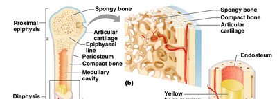
Long bones are characterized by a central shaft and two ends. They contain both compact and spongy bone, and are covered by specialized membranes.
Proximal and distal epiphysis: Ends of the bone, often involved in joint formation
Diaphysis: Shaft of the bone
Medullary cavity: Central cavity containing bone marrow
Epiphyseal line: Remnant of the growth plate
Compact bone: Dense outer layer
Spongy bone: Porous inner structure
Periosteum: Outer fibrous covering
Endosteum: Inner lining of the medullary cavity
Appendicular Skeleton
Pectoral and Pelvic Girdles
The appendicular skeleton includes the limbs and girdles that attach them to the axial skeleton. The pectoral girdle consists of the scapula and clavicle, while the pelvic girdle is formed by the os coxae.
Pectoral girdle:
Scapula: Glenoid cavity, spine, acromion process, coracoid process, borders, angles, fossae, tubercles
Clavicle: Sternal end, acromial end
Pelvic girdle:
Os coxae: Acetabulum, obturator foramen
Ilium: Crest, ala, arcuate line, body, sciatic notch, fossa, spines
Ischium: Tuberosity, spine, sciatic notch
Pubis: Ramus, symphysis, arch
Upper and Lower Appendages
The limbs are composed of multiple bones, each with distinct features for muscle attachment and articulation.
Upper appendage: Humerus, radius, ulna, carpals, metacarpals, phalanges
Lower appendage: Femur, patella, tibia, fibula, tarsals, metatarsals, phalanges
Articulations (Joints)
Knee and Shoulder Joints
Joints are sites where bones meet and allow movement. The knee and shoulder are complex synovial joints with multiple supporting structures.
Knee: Quadriceps tendon, patellar ligament, collateral ligaments, cruciate ligaments, menisci
Shoulder: Labrum, coracoacromial ligament, subacromial bursa, fibrous capsule, tendon sheath, articular cartilage, biceps brachii tendon, synovial cavity
Muscle Actions
Types of Movements
Muscles produce a variety of movements at joints, classified by their direction and effect.
Flexion/extension (hyperextension)
Abduction/adduction
Supination/pronation
Protraction/retraction
Dorsiflexion/plantarflexion
Inversion/eversion
Gliding
Rotation (medial/lateral/right/left)
Circumduction
Opposition
Elevation/depression
Muscles of the Head and Neck
Identification, Origin, Insertion, and Action
Muscles of the head and neck are responsible for facial expressions, mastication, and movement of the head. Each muscle has a specific origin (less movable attachment), insertion (more movable attachment), and action.
Muscles of the scalp: Epicranius (occipitofrontalis)
Muscles of the mouth: Buccinator, levator labii superioris, depressor labii inferioris, depressor anguli oris*, mentalis, orbicularis oris, zygomaticus*
Muscles of the eye: Orbicularis oculi
Muscles of the neck: Platysma
Muscles of mastication: Temporalis*, masseter*, medial pterygoid*, lateral pterygoid
Muscles of mandibular depression: Digastric*
Muscles that move the head & neck: Sternocleidomastoid*, scalene*, splenius*
Muscle | Origin | Insertion | Action |
|---|---|---|---|
Epicranius (occipitofrontalis) | NR | NR | Raises eyebrows, wrinkles forehead |
Buccinator | NR | NR | Compresses cheek |
Orbicularis oris | NR | NR | Closes and puckers lips |
Zygomaticus* | NR | NR | Smiling |
Orbicularis oculi | NR | NR | Blinks, closes eye |
Platysma | NR | NR | Tenses skin of neck |
Temporalis* | NR | NR | Elevates mandible |
Masseter* | NR | NR | Elevates mandible |
Digastric* | NR | NR | Depresses mandible |
Sternocleidomastoid* | NR | NR | Rotates and flexes neck |
Additional info:
NR = Not Recorded in provided materials; refer to textbook for detailed origin and insertion.
Muscles marked with * have antagonists listed.
Muscle actions are essential for understanding movement and clinical assessment of injuries.
