 Back
BackStudy Guide: Bone Anatomy and Physiology (Skeletal System)
Study Guide - Smart Notes
 Tailored notes based on your materials, expanded with key definitions, examples, and context.
The Skeletal System: Overview
Functions of the Skeletal System
The skeletal system is a vital organ system that provides structural support, protection, movement, and storage for the human body. It consists of bones, joints, cartilages, and ligaments.
Protection: Shields internal organs such as the brain, heart, and lungs.
Storage: Stores fats and minerals, especially calcium and phosphate (98% of body Ca2+).
Blood Cell Production: Red bone marrow produces red blood cells, white blood cells, and platelets (hematopoiesis).
Movement: Provides attachment points for muscles, enabling movement.
Structural Support: Framework for soft tissues and organs.
Divisions of the Skeletal System
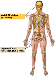
The adult skeleton contains 206 bones, organized into two main divisions:
Axial Skeleton: 80 bones, including the skull, vertebral column, and rib cage.
Appendicular Skeleton: 126 bones, including limbs and girdles.
Classification of Bones
Types of Bone Tissue
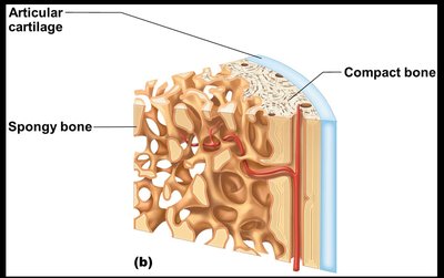
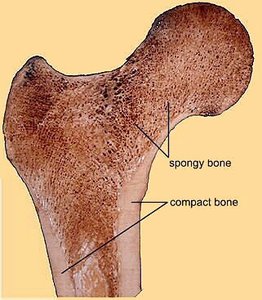
Bones are composed of two main types of osseous tissue:
Compact Bone: Dense, smooth, and homogeneous.
Spongy Bone: Small needlelike pieces with many open spaces.
Bone Shapes and Examples
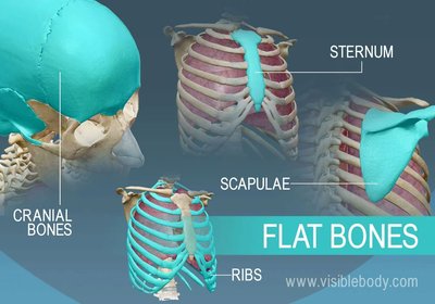
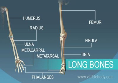
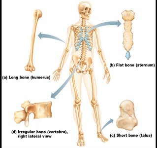
Bones are classified by shape into four main categories:
Flat Bones: Thin, flattened, usually curved. Example: skull, sternum, ribs, scapulae.
Long Bones: Longer than wide, shaft with enlarged ends. Example: femur, humerus.
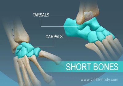
Short Bones: Cube-shaped, mostly spongy bone. Example: carpals, tarsals, patella (sesamoid).
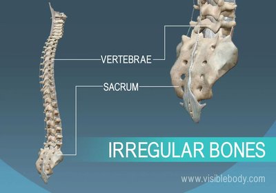
Irregular Bones: Complex shapes. Example: vertebrae, hip bones.

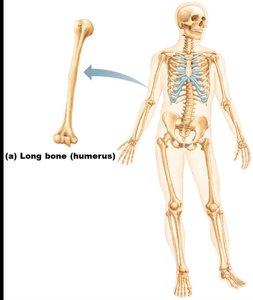
Long Bone Anatomy
Gross Anatomy of a Long Bone
Long bones have a distinct structure with specialized regions:
Diaphysis: Shaft, composed of compact bone.
Epiphysis: Ends, mostly spongy bone enclosed by compact bone.
Metaphysis: Region connecting epiphysis to diaphysis.
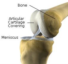
Articular Cartilage: Covers epiphyses, made of hyaline cartilage, reduces friction and absorbs shock.
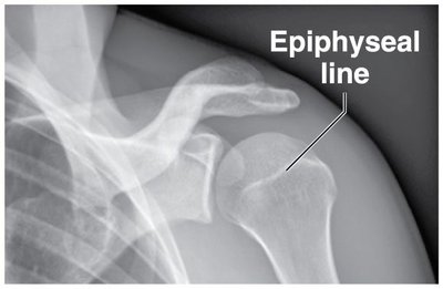
Epiphyseal Plate/Line: Plate is hyaline cartilage in growing bones; line is its remnant in adults.
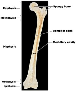
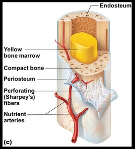
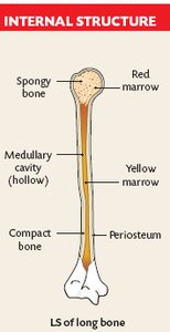
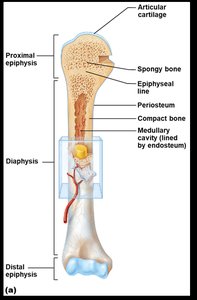
Medullary Cavity and Bone Marrow
Medullary Cavity: Central cavity in diaphysis, contains bone marrow.
Bone Marrow:
Yellow Marrow: Fat storage, found in adults.
Red Marrow: Blood cell formation, found in children.
Bone Cells
Types of Bone Cells
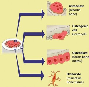
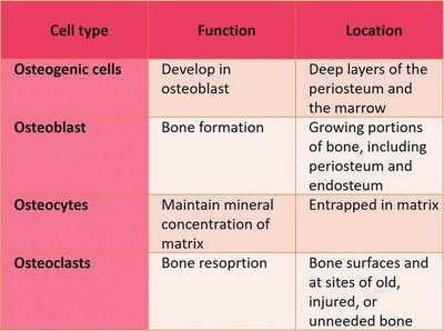
Four main types of bone cells are involved in bone maintenance and repair:
Cell type | Function | Location |
|---|---|---|
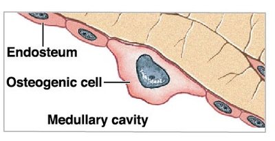
Osteogenic cells | Develop into osteoblasts | Deep layers of periosteum and marrow |
Osteoblasts | Bone formation | Growing portions of bone, periosteum, endosteum |
Osteocytes | Maintain mineral concentration of matrix | Entrapped in matrix |
Osteoclasts | Bone resorption | Bone surfaces, sites of old/injured bone |
Cell Functions
Osteogenic Cells: Stem cells, differentiate into osteoblasts, important for fracture repair.
Osteoblasts: Produce new bone matrix (osteoid), initiate ossification, become osteocytes when surrounded by matrix.
Osteocytes: Mature bone cells, maintain matrix, occupy lacunae, interconnected by canaliculi.
Osteoclasts: Multinucleated, resorb bone matrix, release stored minerals, derived from monocyte lineage.
Microscopic Anatomy of Bone
Compact Bone
Osteon (Haversian System): Basic unit, concentric lamellae, central canal with blood vessels and nerves.
Lacunae: Cavities housing osteocytes.
Canaliculi: Tiny canals connecting lacunae and central canal.
Perforating (Volkmann's) Canals: Connect osteons, carry blood vessels and nerves.
Spongy Bone
Trabeculae: Network of struts and plates, porous, reduces weight, contains bone marrow.
Open Spaces: Filled with marrow, blood vessels, and nerves.
Bone Formation, Growth, and Remodeling
Ossification
Ossification: Process of bone formation, occurs on hyaline cartilage models or fibrous membranes.
Long Bone Growth: Two phases:
Osteoblasts cover cartilage model with bone matrix.
Cartilage is digested away, opening medullary cavity.
Appositional Growth
Appositional Growth: Increase in bone diameter, osteoblasts add matrix to outside, osteoclasts remove matrix from inside.
Controlled by Hormones: Growth hormone and sex hormones regulate bone growth.
Bone Remodeling
Remodeling: Occurs throughout life, regulated by calcium ion levels and mechanical stress.
Calcium Regulation:
Parathyroid Hormone (PTH): Increases osteoclast activity, releases calcium.
Calcitonin: Decreases osteoclast activity, increases calcium storage.
Fracture and Repair
Types of Fractures
Closed (Simple) Fracture: Does not penetrate skin.
Open (Compound) Fracture: Penetrates skin, risk of infection.
Fracture Repair Steps
Fracture Hematoma: Blood-filled swelling forms.
Fibrocartilage Callus Formation: Cartilage and collagen splint the bone.
Bony Callus Formation: Osteoblasts and osteoclasts migrate in.
Bone Remodeling: Bone reshaped in response to stress.
Normal Aging and Osteoporosis
Normal Aging
Bones become thinner and weaker after age 30–40.
Osteoblast activity declines, osteoclast activity remains.
Women lose more skeletal mass per decade than men.
Osteoporosis
Loss of bone mass compromises function.
Increases risk of fractures, especially in vertebrae.
Summary Table: Bone Cell Types
Cell type | Function | Location |
|---|---|---|
Osteogenic cells | Develop into osteoblasts | Deep layers of periosteum and marrow |
Osteoblasts | Bone formation | Growing portions of bone, periosteum, endosteum |
Osteocytes | Maintain mineral concentration of matrix | Entrapped in matrix |
Osteoclasts | Bone resorption | Bone surfaces, sites of old/injured bone |
Self-Check Questions
Describe the functions of the skeletal system.
Classify bones according to their shapes, and give examples of each type.
State the locations of compact and spongy bone and compare their structures and functions.
Identify the parts of a typical long bone and describe its internal anatomy.
Name the three cell types in bone, identify their major functions in bone maintenance and healing.
Describe the processes of bone development, growth, and remodeling.
Describe the changes between epiphyseal plate vs line.
Summarize calcium homeostasis.
Identify bone fracture types, and summarize the repair steps.
