 Back
BackStudy Guide: Bones and Bone Markings of the Human Skeleton
Study Guide - Smart Notes
 Tailored notes based on your materials, expanded with key definitions, examples, and context.
Bones and Bone Markings of the Human Skeleton
Overview of the Human Skeleton
The human skeleton is divided into two main regions: the axial skeleton and the appendicular skeleton. Each region contains specific bones and markings essential for structure, movement, and protection.
Axial Skeleton: Includes the skull, vertebral column, and thoracic cage. Provides support and protection for the brain, spinal cord, and thoracic organs.
Appendicular Skeleton: Comprises the limbs and girdles, facilitating movement and interaction with the environment.
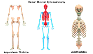
Axial Skeleton
Skull
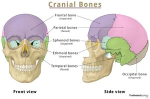
The skull consists of cranial and facial bones, which protect the brain and form the structure of the face.
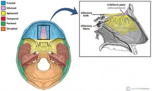
Cranial Bones: Frontal (unpaired), parietal (paired), sphenoid (unpaired), ethmoid (unpaired), temporal (paired), occipital (unpaired).
Sutures: Immovable joints connecting cranial bones.
Key Markings:
Cribriform plate (ethmoid): Contains foramina for olfactory nerves.
Mastoid process (temporal): Attachment for neck muscles.
Foramen magnum (occipital): Passage for spinal cord.
Facial Bones
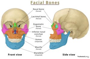
Facial bones form the structure of the face and support sensory organs.
Nasal, lacrimal, zygomatic, inferior nasal conchae, vomer, maxilla, mandible
Key Markings:
Alveolar processes (maxilla): Hold teeth.
Condylar process (mandible): Forms the temporomandibular joint (TMJ).
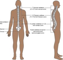
Vertebral Column
The vertebral column supports the body and protects the spinal cord.
Cervical vertebrae (7): C1 (Atlas), C2 (Axis, with dens).
Thoracic vertebrae (12)
Lumbar vertebrae (5): Body, vertebral foramen, intervertebral foramina.
Sacrum: 3-4 fused vertebrae.
Coccyx: Terminal portion.
Thoracic Cage
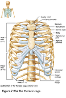
The thoracic cage protects vital organs and supports respiration.
Ribs (12):
True ribs (1-7): Directly attached to sternum.
False ribs (8-12): Indirectly or not attached to sternum.
Floating ribs (11-12): No anterior attachment.
Sternum: Manubrium, body, xiphoid process; jugular and clavicular notches.
Costal cartilage: Connects ribs to sternum.
Appendicular Skeleton
Pectoral Girdle and Upper Limb
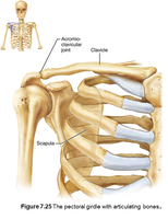
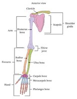
The pectoral girdle anchors the upper limbs to the axial skeleton.
Clavicle: Acromial and sternal ends.
Scapula: Acromial clavicular joint, glenoid cavity.
Humerus: Head, tubercles, trochlea, capitulum.
Radius: Head, styloid process.
Ulna: Olecranon, styloid process.
Carpals (8), metacarpals, phalanges (proximal, intermediate, distal; pollex lacks intermediate).
Pelvic Girdle and Lower Limb
The pelvic girdle supports the lower limbs and protects pelvic organs.
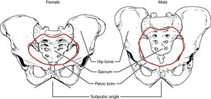
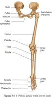
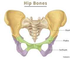
Coxal bones: Ilium, pubis, ischium; sacroiliac joints, pubic symphysis, pubic brim & arch (male vs. female).
Femur: Head, medial and lateral condyles.
Patella: Kneecap.
Tibia: Medial and lateral condylar surfaces (tibial plateau & meniscus).
Fibula: Lateral bone of the leg.
Tarsals (7): Includes calcaneus (heel bone).
Metatarsals, phalanges (proximal, intermediate, distal; hallux lacks intermediate).
Calcaneus Bone
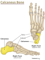
The calcaneus is the largest tarsal bone and forms the heel.
Function: Supports body weight and provides attachment for the Achilles tendon.
Location: Posterior part of the foot, articulates with the talus and cuboid bones.
Summary Table: Major Bones of the Human Skeleton
Region | Main Bones | Key Markings/Features |
|---|---|---|
Skull | Frontal, parietal, sphenoid, ethmoid, temporal, occipital, nasal, lacrimal, zygomatic, maxilla, mandible | Sutures, cribriform plate, mastoid process, foramen magnum, alveolar processes, condylar process |
Vertebral Column | Cervical (C1, C2), thoracic, lumbar, sacrum, coccyx | Dens, vertebral foramen, intervertebral foramina |
Thoracic Cage | Ribs, sternum, costal cartilage | True/false/floating ribs, manubrium, jugular notch |
Pectoral Girdle | Clavicle, scapula | Acromial/sterna ends, glenoid cavity |
Upper Limb | Humerus, radius, ulna, carpals, metacarpals, phalanges | Tubercles, trochlea, capitulum, olecranon, styloid process |
Pelvic Girdle | Ilium, pubis, ischium, sacrum, coccyx | Sacroiliac joint, pubic symphysis, pubic brim/arch |
Lower Limb | Femur, patella, tibia, fibula, tarsals, metatarsals, phalanges | Condyles, tibial plateau, meniscus, calcaneus |
Additional info:
The pollex (thumb) and hallux (big toe) lack intermediate phalanges.
Male and female pelvic brims differ in shape and subpubic angle, affecting childbirth.
