Back
Study Guide: Bones and Features of the Human Skeleton (ANP College Level)
Study Guide - Smart Notes
 Tailored notes based on your materials, expanded with key definitions, examples, and context.
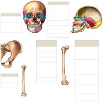
Q1. Identify and describe the major openings, elevations, and depressions found in bones of the human skeleton.
Background
Topic: Bone Markings and Features
This question tests your knowledge of anatomical terminology for bone features, including openings (foramina, canals), elevations (processes, tubercles), and depressions (fossae, sulci). Understanding these terms is essential for identifying bones and their functions in the skeletal system.
Key Terms:
Foramen: An opening in a bone for blood vessels or nerves.
Process: A projection or bump on a bone.
Fossa: A shallow depression.
Canal/Meatus: Passage or channel within a bone.
Tubercle/Tuberosity: Rough projections for muscle attachment.
Condyle: Smooth, rounded articular surface.
Spine: Pointed process.
Step-by-Step Guidance
Review the definitions of each bone feature and consider their functional significance (e.g., foramen allows passage of nerves).
Examine diagrams of the skull, pelvis, and long bones to locate these features visually.
Match each term to its corresponding location on a bone (e.g., foramen magnum in the skull, greater trochanter on the femur).
Think about how these features relate to muscle attachment, joint formation, or passage of structures.
Try solving on your own before revealing the answer!
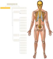
Q2. Classify the bones of the human skeleton into axial and appendicular divisions.
Background
Topic: Skeletal System Organization
This question assesses your understanding of the two main divisions of the skeleton: axial (skull, vertebral column, rib cage) and appendicular (limbs and girdles).
Key Terms:
Axial Skeleton: Includes skull, vertebrae, ribs, and sternum.
Appendicular Skeleton: Includes limbs, pectoral and pelvic girdles.
Step-by-Step Guidance
Recall the definition and components of the axial skeleton.
Recall the definition and components of the appendicular skeleton.
Use a diagram of the full skeleton to visually separate the two divisions.
List examples of bones in each division (e.g., femur in appendicular, vertebrae in axial).
Try solving on your own before revealing the answer!
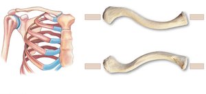
Q3. Identify the anatomical features of the clavicle and their functional significance.
Background
Topic: Pectoral Girdle Anatomy
This question focuses on the structure of the clavicle, including its ends, tubercles, and articulations, and their roles in shoulder stability and movement.
Key Terms:
Acromial End: Lateral end articulating with scapula.
Sternal End: Medial end articulating with sternum.
Conoid Tubercle: Attachment for ligaments.
Costal Tuberosity: Attachment for ligaments.
Step-by-Step Guidance
Identify the two ends of the clavicle and their articulations.
Locate the conoid tubercle and costal tuberosity on diagrams.
Consider how these features contribute to shoulder stability.
Relate the anatomical features to their functional roles (e.g., ligament attachment).
Try solving on your own before revealing the answer!
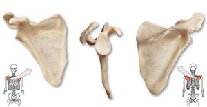
Q4. Describe the anatomical features of the scapula and their importance in shoulder movement.
Background
Topic: Scapular Anatomy
This question tests your ability to identify the major landmarks of the scapula and understand their roles in muscle attachment and joint formation.
Key Terms:
Acromion: Articulates with clavicle.
Coracoid Process: Muscle attachment.
Glenoid Cavity: Articulates with humerus.
Spine: Divides the scapula into fossae.
Step-by-Step Guidance
Identify the acromion, coracoid process, and glenoid cavity on diagrams.
Locate the spine and fossae (supraspinous, infraspinous, subscapular).
Relate each feature to its function (e.g., muscle attachment, joint formation).
Consider how the scapula's features enable shoulder movement.
Try solving on your own before revealing the answer!
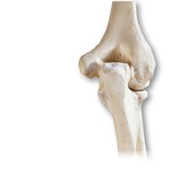
Q5. Identify the bones and features of the elbow joint.
Background
Topic: Elbow Joint Anatomy
This question focuses on the bones (humerus, radius, ulna) and their features that form the elbow joint, including articulating surfaces and processes.
Key Terms:
Olecranon: Process of ulna forming the elbow.
Trochlea: Articulates with ulna.
Capitulum: Articulates with radius.
Coronoid Process: Part of ulna.
Step-by-Step Guidance
Identify the humerus, radius, and ulna in the elbow joint diagram.
Locate the olecranon, trochlea, and capitulum.
Understand how these features articulate to allow movement.
Relate the anatomical features to their functional roles in flexion and extension.
Try solving on your own before revealing the answer!
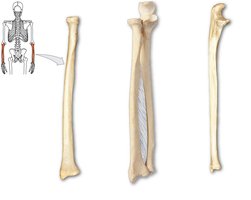
Q6. Describe the anatomical features of the radius and ulna and their relationship in the forearm.
Background
Topic: Forearm Anatomy
This question tests your knowledge of the radius and ulna, their features, and how they interact to allow forearm movement.
Key Terms:
Radial Head: Articulates with humerus.
Olecranon: Process of ulna.
Interosseous Membrane: Connects radius and ulna.
Styloid Process: Distal end of both bones.
Step-by-Step Guidance
Identify the radius and ulna in the forearm diagram.
Locate the radial head, olecranon, and styloid processes.
Understand the role of the interosseous membrane.
Relate the anatomical features to their function in pronation and supination.
Try solving on your own before revealing the answer!
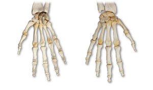
Q7. Identify the bones of the hand and their arrangement.
Background
Topic: Hand Anatomy
This question focuses on the arrangement of carpal, metacarpal, and phalangeal bones in the hand.
Key Terms:
Carpals: Wrist bones.
Metacarpals: Palm bones.
Phalanges: Finger bones.
Step-by-Step Guidance
Identify the carpal, metacarpal, and phalangeal bones in the hand diagram.
Count the number of each type of bone.
Understand the arrangement and articulation of these bones.
Relate the anatomical arrangement to hand function and movement.
Try solving on your own before revealing the answer!
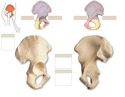
Q8. Identify the anatomical features of the coxal bone and their significance in pelvic structure.
Background
Topic: Pelvic Girdle Anatomy
This question tests your knowledge of the coxal bone (ilium, ischium, pubis) and its features, including the acetabulum and obturator foramen.
Key Terms:
Ilium: Superior part of coxal bone.
Ischium: Posterior part.
Pubis: Anterior part.
Acetabulum: Hip socket.
Obturator Foramen: Large opening.
Step-by-Step Guidance
Identify the ilium, ischium, and pubis in the coxal bone diagram.
Locate the acetabulum and obturator foramen.
Understand the significance of these features in weight-bearing and movement.
Relate the anatomical features to pelvic structure and function.
Try solving on your own before revealing the answer!
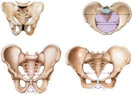
Q9. Compare the male and female pelvis and describe their differences.
Background
Topic: Pelvic Anatomy and Sexual Dimorphism
This question focuses on the structural differences between male and female pelvises, including pelvic inlet, outlet, and angle.
Key Terms:
Pelvic Inlet/Outlet: Openings in the pelvis.
Subpubic Angle: Angle beneath pubic symphysis.
False/True Pelvis: Divisions of the pelvis.
Step-by-Step Guidance
Identify the pelvic inlet, outlet, and subpubic angle in diagrams.
Compare the shape and size of male and female pelvises.
Understand the functional significance (e.g., childbirth).
Relate anatomical differences to clinical or functional outcomes.
Try solving on your own before revealing the answer!
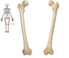
Q10. Identify the anatomical features of the femur and their functional significance.
Background
Topic: Femur Anatomy
This question tests your knowledge of the femur's features, including head, neck, trochanters, and condyles, and their roles in movement and weight-bearing.
Key Terms:
Femoral Head: Articulates with pelvis.
Neck: Connects head to shaft.
Greater/Lesser Trochanter: Muscle attachment.
Condyles: Articulate with tibia.
Step-by-Step Guidance
Identify the head, neck, and trochanters in the femur diagram.
Locate the condyles and relate them to knee articulation.
Understand the functional significance of each feature.
Relate anatomical features to movement and weight-bearing.
Try solving on your own before revealing the answer!
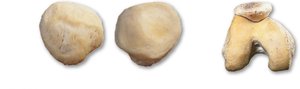
Q11. Describe the anatomical features of the patella and its role in the knee joint.
Background
Topic: Patella Anatomy
This question focuses on the patella's features and its function in protecting the knee and improving leverage for the quadriceps muscle.
Key Terms:
Base: Superior part.
Apex: Inferior part.
Articular Surface: Contacts femur.
Step-by-Step Guidance
Identify the base, apex, and articular surface in the patella diagram.
Understand the patella's role in knee extension.
Relate anatomical features to function and protection.
Consider how the patella improves leverage for the quadriceps.
Try solving on your own before revealing the answer!
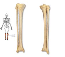
Q12. Identify the anatomical features of the tibia and fibula and their relationship in the lower leg.
Background
Topic: Lower Leg Anatomy
This question tests your knowledge of the tibia and fibula, their features, and how they interact to support weight and allow movement.
Key Terms:
Tibial Tuberosity: Attachment for patellar ligament.
Medial/Lateral Malleolus: Ankle prominences.
Interosseous Membrane: Connects tibia and fibula.
Step-by-Step Guidance
Identify the tibia and fibula in the lower leg diagram.
Locate the tibial tuberosity and malleoli.
Understand the role of the interosseous membrane.
Relate anatomical features to weight-bearing and movement.
Try solving on your own before revealing the answer!
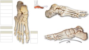
Q13. Identify the bones of the foot and their arrangement.
Background
Topic: Foot Anatomy
This question focuses on the arrangement of tarsal, metatarsal, and phalangeal bones in the foot.
Key Terms:
Tarsals: Ankle bones.
Metatarsals: Foot bones.
Phalanges: Toe bones.
Arches: Longitudinal and transverse.
Step-by-Step Guidance
Identify the tarsal, metatarsal, and phalangeal bones in the foot diagram.
Count the number of each type of bone.
Understand the arrangement and articulation of these bones.
Relate the anatomical arrangement to foot function and movement.
Try solving on your own before revealing the answer!