Back
BackStudy Guide: Bones and Skeletal Tissues (Chapter 6)
Study Guide - Smart Notes
 Tailored notes based on your materials, expanded with key definitions, examples, and context.
Q1. Assign each variety of cartilage (hyaline cartilage, elastic cartilage, and fibrocartilage) to the description of its function.
Background
Topic: Types of Cartilage
This question tests your understanding of the functional properties of the three main types of cartilage found in the human body.
Key Terms:
Hyaline cartilage
Elastic cartilage
Fibrocartilage
Step-by-Step Guidance
Read each functional description carefully: maintaining shape with flexibility, resisting compression and providing tensile strength, and providing support with flexibility and resilience.
Recall the main locations and roles of each cartilage type in the body (e.g., ear, joints, intervertebral discs).
Match each cartilage type to the function based on its structural properties and where it is found in the body.
Try solving on your own before revealing the answer!
Q2. Complete the table by assigning each variety of cartilage (hyaline cartilage, elastic cartilage, and fibrocartilage) to its location in the body.
Background
Topic: Cartilage Locations
This question tests your ability to identify where each type of cartilage is found in the adult skeleton.
Key Terms:
Hyaline cartilage: most abundant, found in joints, ribs, nose, etc.
Elastic cartilage: found in structures needing flexibility (ear, epiglottis).
Fibrocartilage: found in areas needing strength and shock absorption (intervertebral discs, menisci).
Step-by-Step Guidance
List the locations given (e.g., ends of bones at joints, external ear, menisci of knee, etc.).
Recall which cartilage type is associated with each location based on its properties.
Assign the correct cartilage type to each location in the table.
Q3. Explain how cartilage grows (appositional vs. interstitial growth).
Background
Topic: Cartilage Growth Mechanisms
This question tests your understanding of the two main ways cartilage can grow: from within (interstitial) or by adding new layers on the outside (appositional).
Key Terms:
Appositional growth: new layers added to the surface.
Interstitial growth: growth from within the tissue.
Step-by-Step Guidance
Define both types of growth in your own words.
Identify which type involves new cartilage forming inside the tissue and which involves new cartilage forming at the edge.
Fill in the blanks with the correct terms.
Try solving on your own before revealing the answer!
Q4. True or False: The hard extracellular matrix of cartilage tissue allows for mitosis.
Background
Topic: Cartilage Structure and Growth
This question checks your understanding of how the structure of cartilage affects its ability to grow and repair.
Key Terms:
Extracellular matrix
Mitosis
Step-by-Step Guidance
Recall what the extracellular matrix is and its properties in cartilage.
Consider whether a hard or flexible matrix would allow for cell division (mitosis).
Decide if the statement is true or false, and if false, correct the underlined word.
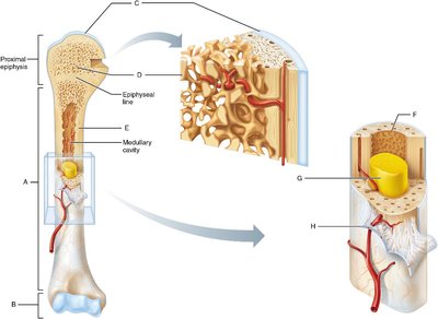
Q5. Indicate the location of the following structures by assigning the letters (A–H) from the figure.
Background
Topic: Gross Anatomy of Long Bone
This question tests your ability to identify anatomical structures of a long bone using a labeled diagram.
Key Terms:
Endosteum
Distal epiphysis
Articular cartilage
Spongy bone
Compact bone
Yellow bone marrow
Periosteum
Diaphysis
Step-by-Step Guidance
Review the labeled diagram of the long bone provided.
Recall the definition and location of each structure listed above.
Match each structure to the correct letter on the diagram.
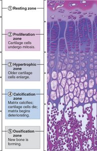
Q6. Assign each of the zones of an epiphyseal plate to their correct locations.
Background
Topic: Epiphyseal Plate Zones
This question tests your understanding of the different zones in the epiphyseal plate and their roles in bone growth.
Key Terms:
Resting zone
Proliferation zone
Hypertrophic zone
Calcification zone
Ossification zone
Step-by-Step Guidance
Study the provided histological image of the epiphyseal plate.
Recall the function of each zone (e.g., cell division, enlargement, calcification, bone formation).
Assign the correct label (A–D) to each zone based on its position and description.
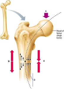
Q7. Assign letters A–D to each of the following conditions represented by the arrows in the femur diagram.
Background
Topic: Mechanical Stress on Bone
This question tests your understanding of how mechanical forces (compression, tension, load, and no stress) are distributed in a long bone such as the femur.
Key Terms:
Compression
Tension
Load application
No stress
Step-by-Step Guidance
Examine the diagram showing the femur and the direction of forces.
Recall where compression and tension typically occur in a loaded bone.
Assign the correct letter to each condition based on the arrows and their locations.