 Back
Study Guide: Classification and Anatomy of Joints (Chapter 8 - ANP College Course)
Study Guide - Smart Notes
 Tailored notes based on your materials, expanded with key definitions, examples, and context.
Joints: Classification and Anatomy
Overview of Joint Classification
Joints, also known as articulations, are connections between bones that allow for varying degrees of movement. They are classified in two main ways: by their anatomical structure and by the range of motion they permit.
Anatomical classification: Based on the material connecting the bones and the presence or absence of a cavity.
Functional classification: Based on the degree of movement allowed at the joint.

Anatomical Ways to Classify Joints
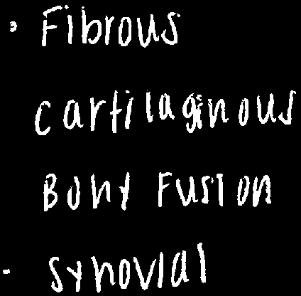
Joints are structurally classified into four main types, each defined by the tissue connecting the bones:
Fibrous joints: Bones are joined by fibrous connective tissue.
Cartilaginous joints: Bones are joined by cartilage.
Bony fusion: Bones are fused together, forming a single structure.
Synovial joints: Bones are separated by a synovial cavity filled with synovial fluid.
Functional Classification of Joints (Range of Motion)
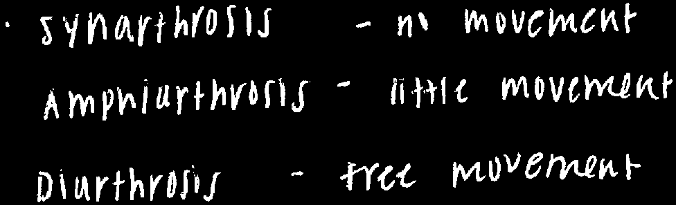
Joints are also classified by the amount of movement they allow:
Synarthrosis: No movement (immovable joints).
Amphiarthrosis: Little movement (slightly movable joints).
Diarthrosis: Free movement (freely movable joints).
Structural Components of Joints
The main structural components found in joints include:
Fibrous connective tissue: Found in fibrous joints, connecting bones directly.
Cartilage: Found in cartilaginous joints, providing cushioning and flexibility.
Synovial cavity: Found in synovial joints, filled with synovial fluid to reduce friction.
Anatomy of a Generic Synovial Joint
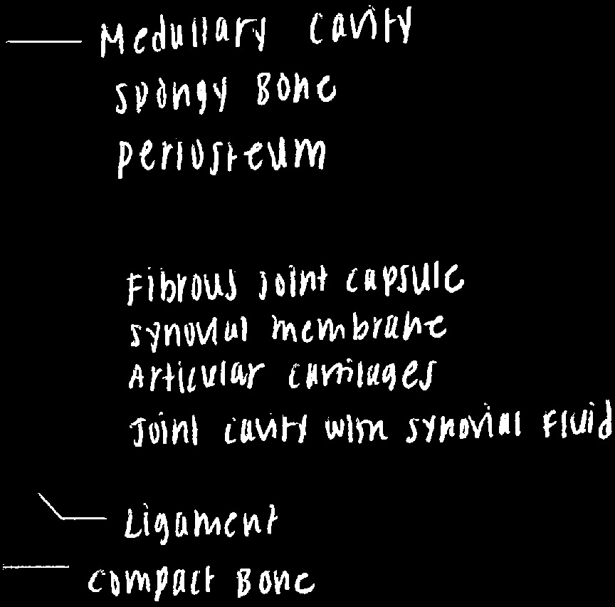
Synovial joints are the most complex and allow the greatest range of movement. Their anatomy includes several key structures:
Articular cartilage: Covers the ends of bones, reducing friction and absorbing shock.
Compact bone: Provides strength and structure.
Fibrous joint capsule: Encloses the joint, providing stability.
Synovial membrane: Lines the joint capsule and produces synovial fluid.
Joint cavity with synovial fluid: Lubricates the joint and nourishes cartilage.
Ligament: Connects bone to bone, stabilizing the joint.
Medullary cavity: Contains bone marrow.
Spongy bone: Provides lightweight support and absorbs shock.
Periosteum: The outer covering of bone.


Functions of Synovial Fluid
Synovial fluid within the joint cavity serves several important functions:
Lubrication and friction reduction: Minimizes wear between articular surfaces.
Nutrient distribution: Supplies nutrients and oxygen to articular cartilage.
Shock absorption: Cushions joints during movement.

Movements Allowed at Synovial Joints
Synovial joints permit a variety of movements, including:
Abduction: Movement away from the midline of the body.
Adduction: Movement toward the midline (opposite of abduction).
Elevation: Moving a structure upward (superiorly).
Depression: Moving a structure downward (inferiorly).
Protraction: Moving a structure anteriorly.
Retraction: Moving a structure posteriorly.
Circumduction: Moving a limb in a circular motion.
Rotation: Turning a bone around its axis (e.g., shaking the head).
Pronation: Turning the palm downward.
Supination: Turning the palm upward.
Eversion: Turning the sole of the foot outward.
Inversion: Turning the sole of the foot inward.

Fibrous Cartilage in Joints
Fibrous cartilage can be present in some joints and serves to limit movement, providing additional stability.
Example: Intervertebral discs are composed of fibrous cartilage and restrict excessive movement between vertebrae.

Bursae
Bursae are small fluid-filled pockets that reduce friction and absorb shock between tendons, ligaments, muscles, and bones.
Function: Facilitate smooth movement and protect tissues from mechanical stress.

Clinical Definitions Related to Joints
Understanding clinical terms is important for recognizing joint injuries and conditions:
Dislocation (luxation): Complete displacement of the bones at a joint, causing loss of contact between articular surfaces.
Inflammation: Swelling, stiffness, pain, and decreased movement due to irritation or injury of the joint.

Summary Table: Joint Classification
The following table summarizes the anatomical and functional classification of joints:
Type | Anatomical Structure | Movement Allowed | Example |
|---|---|---|---|
Fibrous | Fibrous connective tissue | None (synarthrosis) | Sutures of the skull |
Cartilaginous | Cartilage | Little (amphiarthrosis) | Intervertebral discs |
Bony fusion | Bony tissue | None (synarthrosis) | Epiphyseal lines |
Synovial | Synovial cavity with fluid | Free (diarthrosis) | Knee, shoulder |
Additional info:
Some images and notes were inferred to provide complete context for joint anatomy and movement types.
Clinical definitions are essential for understanding joint injuries and pathologies.
