 Back
BackStudy Guide: Epithelial and Connective Tissues (BIO 217)
Study Guide - Smart Notes
 Tailored notes based on your materials, expanded with key definitions, examples, and context.
Types of Tissues in the Human Body
Overview of Tissue Types
Tissues are the fundamental structural and functional units of the human body. Each tissue type is composed of specialized cells and extracellular components, organized to perform specific functions. The four primary tissue types are:
Epithelial tissue: Covers surfaces, lines cavities, and forms glands. Functions include protection, absorption, secretion, and filtration.
Connective tissue: Supports, binds, and protects organs. Characterized by an abundant extracellular matrix. Includes bone, blood, and adipose tissue.
Muscle tissue: Specialized for contraction and movement. Includes skeletal, cardiac, and smooth muscle.
Nervous tissue: Specialized for communication via electrical signals. Includes neurons and supporting neuroglia.
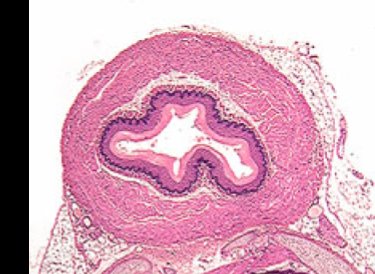
Example: The wall of the esophagus contains all four tissue types, each contributing to its function.
Organization of Tissues in Organ Walls
Layered Structure of Organ Walls
Most organ walls exhibit a layered organization, with each layer composed of a distinct tissue type. The typical arrangement from innermost to outermost is:
Epithelial tissue: Forms the lining (innermost and outermost layers).
Basement membrane: Anchors epithelium to underlying connective tissue.
Connective tissue: Provides structural support, contains blood vessels, nerves, and lymphatics.
Muscle tissue: Responsible for movement and contraction within organ walls.
Example: Hollow organs such as blood vessels, stomach, and intestines follow this pattern.
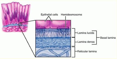
Epithelial Tissue
Structure of Epithelial Tissue
Epithelial tissue is characterized by tightly packed cells with distinct surfaces:
Apical surface: Faces the lumen or external environment.
Basal surface: Attached to the basement membrane.
Basement membrane: Acellular structure composed of proteins and fibers, consisting of two layers:
Basal lamina: Produced by epithelial cells (includes lamina lucida and lamina densa).
Reticular lamina: Produced by underlying connective tissue.
Characteristics of Epithelial Tissue
Polarity: Distinct apical and basal surfaces.
Specialized contacts: Tight junctions and desmosomes form strong barriers.
Supported by connective tissue: Anchored by basement membrane.
Avascular but innervated: No blood vessels; nutrients diffuse from underlying connective tissue.
Regeneration: High mitotic rate allows for rapid repair.
Classification of Epithelia
Epithelia are classified based on cell layers and cell shape:
Number of cell layers:
Simple: One layer
Stratified: Multiple layers
Pseudostratified: Appears multilayered but is a single layer
Cell shapes:
Squamous: Flat, scale-like
Cuboidal: Cube-shaped
Columnar: Tall, elongated
Transitional: Shape changes with tissue stretch (found in urinary tract)
Cell modifications: Ciliated, keratinized, etc.
Examples of Epithelial Tissues
Simple squamous epithelium: Found in lungs; allows for rapid diffusion.
Simple cuboidal epithelium: Found in kidneys; involved in secretion and absorption.
Simple columnar epithelium: Found in small intestine; contains goblet cells for mucus secretion.
Pseudostratified columnar epithelium: Found in trachea; ciliated, contains goblet cells.
Stratified squamous epithelium: Found in skin and esophagus; provides protection.
Connective Tissue
Common Characteristics of Connective Tissues
Common origin: Derived from embryonic mesenchyme.
Vascularity: Most are vascular; exceptions include cartilage and dense regular connective tissue.
Cell dispersion: Cells are widely separated by extracellular matrix.
Location: Found within the body, never exposed to external environment.
Extracellular matrix: Composed of ground substance (glycoproteins, minerals) and fibers (collagen, elastic, reticular).
Classification of Connective Tissue
Connective Tissue Proper:
Loose connective tissue: Loosely arranged fibers; includes areolar and adipose tissue.
Dense connective tissue: Densely packed fibers; includes tendons, ligaments, and dermis.
Specialized Connective Tissue:
Supporting connective tissue: Cartilage and bone.
Fluid connective tissue: Blood and lymph.
Examples of Connective Tissue
Areolar tissue: Holds organs in place; contains collagen, elastin, and reticular fibers.
Adipose tissue: Stores energy, provides cushioning and insulation; nucleus pushed to cell membrane by lipid vacuole.
Dense regular connective tissue: Found in tendons; thick, parallel collagen fibers.
Dense irregular connective tissue: Found in dermis; collagen fibers arranged irregularly.
Hyaline cartilage: Found in trachea; chondrocytes in lacunae within matrix.
Blood: Plasma is the matrix; contains erythrocytes and leukocytes.
Portfolio Assignment for Lab
Field Guide to Connective Tissue
Students are required to document and describe connective tissue types observed in lab, using their own photos and explanations. Assignment includes descriptions and labeled photos of areolar, adipose, dense regular, dense irregular, blood, and hyaline cartilage tissues.
Summary Table: Classification of Epithelial Tissue
Layer Type | Cell Shape | Location | Function |
|---|---|---|---|
Simple | Squamous | Lung alveoli | Diffusion |
Simple | Cuboidal | Kidney tubules | Secretion, absorption |
Simple | Columnar | Small intestine | Absorption, secretion |
Pseudostratified | Columnar | Trachea | Secretion, movement of mucus |
Stratified | Squamous | Skin, esophagus | Protection |
Transitional | Variable | Urinary tract | Stretch |
Summary Table: Classification of Connective Tissue
Class | Type | Location | Function |
|---|---|---|---|
Connective Tissue Proper | Areolar | Under epithelia | Support, binding |
Connective Tissue Proper | Adipose | Subcutaneous tissue | Energy storage, insulation |
Connective Tissue Proper | Dense regular | Tendons, ligaments | Strength, attachment |
Connective Tissue Proper | Dense irregular | Dermis | Strength in multiple directions |
Specialized | Hyaline cartilage | Trachea, joints | Support, flexibility |
Specialized | Blood | Blood vessels | Transport |
Additional info: Academic context was added to clarify tissue functions, structural characteristics, and examples. Tables were inferred and expanded for completeness.
