 Back
BackStudy Guide for BIO163: Endocrine and Special Senses (Eye & Ear)
Study Guide - Smart Notes
 Tailored notes based on your materials, expanded with key definitions, examples, and context.
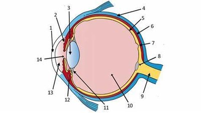
Q1. Identify and describe the structures of the eye in the diagram. For each structure, list its physiology/function and location (chamber/layer).
Background
Topic: Anatomy and Physiology of the Eye
This question tests your knowledge of the anatomical parts of the eye, their physiological roles, and their locations within the eye's chambers or layers.
Key Terms:
Anterior chamber, posterior chamber, sclera, cornea, retina, lens, optic nerve, vitreous body, choroid, ciliary body, iris, pupil, macula, fovea.
Physiology: Vision, light refraction, image formation, signal transmission.
Layers: Fibrous (outer), vascular (middle), neural (inner).
Step-by-Step Guidance
Examine the diagram and identify each numbered structure. Use your textbook or lecture notes to match the numbers to anatomical terms.
For each structure, write a brief description of its function (e.g., "The cornea refracts light entering the eye").
Determine the location of each structure: Is it in the anterior chamber, posterior chamber, or which layer (fibrous, vascular, neural)?
Organize your answers in a table format for clarity, listing structure, function, and location.
Try solving on your own before revealing the answer!
Final Answer:
Each structure corresponds to a specific part of the eye, such as the cornea, lens, retina, etc. Their functions include light refraction, image formation, and signal transmission. Locations are divided among the eye's chambers and layers.
For example, the cornea is in the fibrous layer and anterior chamber, and its function is to refract light. The retina is in the neural layer and posterior chamber, responsible for photoreception.
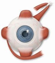
Q2. Label the external eye muscles in the diagram, describe their function, and state their innervation.
Background
Topic: Extraocular Muscles and Eye Movement
This question assesses your understanding of the muscles controlling eye movement, their actions, and the cranial nerves that innervate them.
Key Terms:
Extraocular muscles: superior rectus, inferior rectus, lateral rectus, medial rectus, superior oblique, inferior oblique.
Innervation: Cranial nerves III (oculomotor), IV (trochlear), VI (abducens).
Function: Eye movement in various directions.
Step-by-Step Guidance
Identify each muscle in the diagram and label it with its anatomical name.
For each muscle, describe its primary action (e.g., "The lateral rectus abducts the eye").
List the cranial nerve responsible for innervating each muscle.
Organize your answers in a table for clarity: muscle, function, innervation.
Try solving on your own before revealing the answer!
Final Answer:
The six extraocular muscles control eye movement. Most are innervated by cranial nerve III, except the lateral rectus (VI) and superior oblique (IV). Each muscle moves the eye in a specific direction.
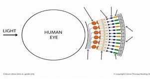
Q3. Label the photoreceptors in the diagram and list the function of each.
Background
Topic: Photoreceptors and Visual Processing
This question tests your knowledge of the types of photoreceptors in the retina and their roles in vision.
Key Terms:
Photoreceptors: rods and cones.
Function: Rods detect dim light and are responsible for night vision; cones detect color and are responsible for high-acuity vision.
Step-by-Step Guidance
Identify the two main types of photoreceptors in the diagram: rods and cones.
Label each photoreceptor and describe its function (e.g., "Rods are sensitive to low light levels").
Explain how the distribution of rods and cones affects vision (e.g., cones are concentrated in the fovea).
Try solving on your own before revealing the answer!
Final Answer:
Rods are responsible for night vision and are more numerous in the peripheral retina. Cones are responsible for color vision and visual acuity, concentrated in the fovea.
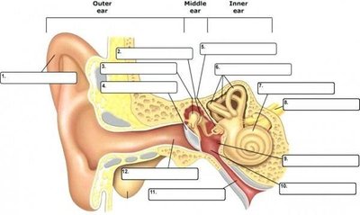
Q4. Label all structures in the ear diagram and list the function of each. Know the ossicles in order, organs for static/dynamic equilibrium, hearing sensation, and VIIIth cranial nerve divisions and function.
Background
Topic: Anatomy and Physiology of the Ear
This question covers the structure and function of the ear, including hearing and balance, and the role of the vestibulocochlear nerve (cranial nerve VIII).
Key Terms:
Ossicles: malleus, incus, stapes (in order).
Organs: cochlea (hearing), vestibule (static equilibrium), semicircular canals (dynamic equilibrium).
Cranial nerve VIII: cochlear division (hearing), vestibular division (balance).
Step-by-Step Guidance
Label each structure in the diagram: outer ear, middle ear, inner ear, ossicles, cochlea, vestibule, semicircular canals.
List the function of each structure (e.g., "The cochlea converts sound waves into nerve impulses").
Identify the order of the ossicles and their role in sound transmission.
Describe which organs are responsible for static and dynamic equilibrium, and hearing sensation.
Explain the divisions and functions of cranial nerve VIII.
Try solving on your own before revealing the answer!
Final Answer:
The ossicles are malleus, incus, and stapes. The cochlea is for hearing, vestibule for static equilibrium, semicircular canals for dynamic equilibrium. Cranial nerve VIII has cochlear (hearing) and vestibular (balance) divisions.
