 Back
BackStudy Guide for Sensory System: Anatomy & Physiology I (BIOL 2401)
Study Guide - Smart Notes
 Tailored notes based on your materials, expanded with key definitions, examples, and context.
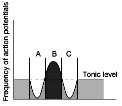
Q27. Consider this diagram showing lateral inhibition and answer the questions:
Background
Topic: Sensory Physiology – Lateral Inhibition
This question tests your understanding of how lateral inhibition enhances sensory acuity by inhibiting neighboring neurons, allowing the nervous system to better localize stimuli.
Key Terms and Concepts:
Lateral inhibition: A process where excited neurons reduce the activity of neighboring neurons, sharpening sensory perception.
Neurotransmitter (NT): Chemical messengers released by neurons to transmit signals.
Secondary neurons: Neurons that receive input from primary sensory neurons.
Step-by-Step Guidance
Examine the diagram: It shows three neurons (A, B, C) with varying frequencies of action potentials. The central neuron (B) has the highest frequency, indicating it received the strongest stimulus.
Identify which neurons are inhibited: Notice the dips in action potential frequency for neurons A and C compared to B. This suggests lateral inhibition is occurring.
Determine the inhibiting neuron: The neuron with the highest activity (B) is likely inhibiting its neighbors (A and C) through interneurons.
Consider neurotransmitter release: The neuron with the highest action potential frequency (B) will release the most neurotransmitter at the synapse.
Try solving on your own before revealing the answer!
Final Answer:
Neurons A and C were inhibited. Neuron B is the inhibiting neuron and produced the highest amount of neurotransmitter.
Lateral inhibition increases acuity by suppressing the activity of neighboring neurons, allowing the nervous system to pinpoint the location of the stimulus more precisely.
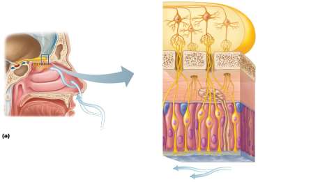
Q24. Label the image with the following terms: olfactory receptor cells, olfactory epithelium, supporting cells, and basal cells.
Background
Topic: Olfaction – Structure of the Olfactory Epithelium
This question tests your ability to identify and label the cellular components of the olfactory epithelium, which is essential for the sense of smell.
Key Terms:
Olfactory receptor cells: Sensory neurons responsible for detecting odorants.
Olfactory epithelium: Specialized tissue in the nasal cavity containing olfactory cells.
Supporting cells: Provide structural and metabolic support to olfactory receptor cells.
Basal cells: Stem cells that regenerate olfactory receptor cells.
Step-by-Step Guidance
Locate the olfactory epithelium in the image: It is the layer lining the upper part of the nasal cavity.
Identify olfactory receptor cells: These are elongated cells extending from the surface of the epithelium to the underlying tissue.
Find supporting cells: These are adjacent to the olfactory receptor cells, often taller and provide support.
Locate basal cells: These are found at the base of the epithelium, near the basement membrane.
Try solving on your own before revealing the answer!
Final Answer:
The olfactory epithelium contains olfactory receptor cells (sensory neurons), supporting cells (structural support), and basal cells (regeneration).
Labeling these structures helps you understand how the sense of smell is maintained and regenerated.
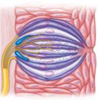
Q28. Label the following structures on the picture of the taste buds below: taste bud, basal cells, gustatory receptor cells, taste pore, and microvilli of gustatory receptor cells.
Background
Topic: Gustation – Structure of Taste Buds
This question tests your ability to identify and label the cellular components of a taste bud, which are essential for the sense of taste.
Key Terms:
Taste bud: Cluster of cells responsible for detecting taste stimuli.
Basal cells: Stem cells that regenerate gustatory cells.
Gustatory receptor cells: Sensory cells that detect taste molecules.
Taste pore: Opening at the surface of the taste bud where tastants enter.
Microvilli: Extensions of gustatory cells that increase surface area for tastant binding.
Step-by-Step Guidance
Identify the taste bud: It is the oval cluster of cells in the image.
Locate basal cells: These are found at the base of the taste bud, near the nerve fibers.
Find gustatory receptor cells: These are elongated cells extending from the base to the taste pore.
Locate the taste pore: The small opening at the top of the taste bud.
Identify microvilli: Fine extensions projecting into the taste pore from gustatory cells.
Try solving on your own before revealing the answer!
Final Answer:
The taste bud contains basal cells (regeneration), gustatory receptor cells (taste detection), a taste pore (entry point), and microvilli (surface area for tastant binding).
Labeling these structures helps clarify how taste is detected and processed.
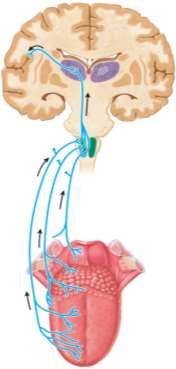
Q30. Label the components of the afferent pathway in the image below.
Background
Topic: Gustatory Pathway – Neural Transmission of Taste
This question tests your ability to identify the neural pathway that transmits taste information from the tongue to the brain.
Key Terms:
Afferent pathway: The route by which sensory information travels to the central nervous system.
Gustatory pathway: The specific neural route for taste sensation.
Brainstem, thalamus, gustatory cortex: Key relay points in the pathway.
Step-by-Step Guidance
Identify the tongue: The origin of the pathway where taste receptor cells are located.
Trace the nerves: Follow the arrows from the tongue to the brainstem.
Locate the thalamus: The relay station in the brain for sensory information.
Find the gustatory cortex: The final destination in the brain where taste is perceived.
Try solving on your own before revealing the answer!
Final Answer:
The afferent pathway for taste includes the tongue, cranial nerves, brainstem, thalamus, and gustatory cortex.
Understanding this pathway is essential for grasping how taste information is processed in the nervous system.
