 Back
BackStudy Guide: Immune, Cardiovascular, and Blood Physiology
Study Guide - Smart Notes
 Tailored notes based on your materials, expanded with key definitions, examples, and context.
Lymphatic System
Organs of the Lymphatic System
The lymphatic system is composed of primary and secondary organs that play crucial roles in immune function and fluid balance.
Primary organs: Sites where lymphocytes are produced and mature (e.g., bone marrow, thymus).
Secondary organs: Sites where immune responses are initiated (e.g., lymph nodes, spleen, tonsils).
Example: The thymus is a primary organ where T cells mature, while lymph nodes are secondary organs where immune cells encounter antigens.
General Function of the Lymphatic System
The lymphatic system maintains fluid balance, absorbs dietary fats, and provides immune defense by transporting lymphocytes and filtering pathogens.
Returns interstitial fluid to the bloodstream
Transports dietary lipids
Facilitates immune surveillance and response
Immune Response
1st Line of Defense: Physical and Chemical Barriers
The first line of defense consists of physical and chemical barriers that prevent pathogen entry.
Skin: Acts as a physical barrier to block pathogens.
Mucus: Traps microbes in respiratory and digestive tracts.
Tears and Saliva: Contain enzymes (e.g., lysozyme) that destroy bacteria.
Other secretions: Such as stomach acid, which kills ingested pathogens.
Example: Tears contain lysozyme, which breaks down bacterial cell walls.
Innate vs Adaptive Immunity
Immunity is divided into innate (nonspecific) and adaptive (specific) responses.
Innate immunity: Immediate, nonspecific defense (e.g., barriers, phagocytes, inflammation).
Adaptive immunity: Delayed, specific response involving lymphocytes (B and T cells) and memory formation.
2nd Line of Defense: Innate Immune Cells and Mechanisms
The second line of defense involves cellular and molecular mechanisms that target pathogens nonspecifically.
White Blood Cells (WBCs): Phagocytes (e.g., neutrophils, macrophages) engulf and destroy pathogens.
Interferons: Proteins that inhibit viral replication.
Inflammation: Local response to injury or infection, increases blood flow and recruits immune cells.
Fever: Systemic increase in body temperature to inhibit pathogens.
Complement System: Plasma proteins that enhance phagocytosis and lyse pathogens.
Phagocytosis: Engulfment and digestion of microbes by phagocytes.
Antigen Presenting Cells (APCs): Present antigens to T cells (e.g., dendritic cells, macrophages).
Natural Killer (NK) Cells: Destroy infected or abnormal cells without prior sensitization.
3rd Line of Defense: Adaptive Immunity
The third line of defense is characterized by specificity and memory, involving B and T lymphocytes.
Humoral Immunity (B cells):
Plasma cells produce antibodies (five classes: IgG, IgA, IgM, IgE, IgD).
Memory B cells provide long-term immunity.
Cell-mediated Immunity (T cells):
Cytotoxic T cells destroy infected cells.
Helper T cells activate other immune cells.
Regulatory T cells modulate immune response.
Memory T cells provide long-term protection.
MHC I vs MHC II: MHC I presents antigens to cytotoxic T cells; MHC II presents to helper T cells.
Immune Response to:
Allergies: Hypersensitivity reactions to harmless antigens.
Bacteria: Phagocytosis and antibody-mediated neutralization.
Viruses: Cytotoxic T cells and interferon response.
NK Cells: Provide rapid response to virally infected cells and tumors.
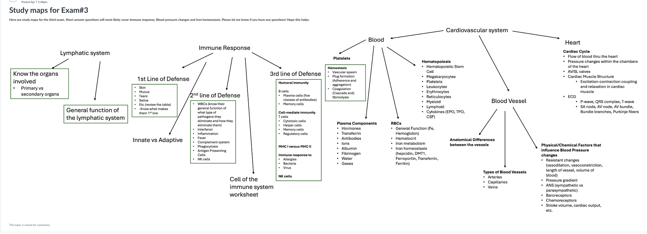
Blood
Hemostasis
Hemostasis is the process that prevents blood loss after vessel injury.
Vascular spasm: Immediate vasoconstriction to reduce blood flow.
Platelet plug formation: Platelets adhere to exposed collagen and aggregate.
Coagulation: Cascade of clotting factors leading to fibrin clot formation.
Fibrinolysis: Breakdown and removal of the clot after healing.
Plasma Components
Plasma is the liquid portion of blood, containing water, proteins, and dissolved substances.
Hormones: Chemical messengers (e.g., insulin, erythropoietin).
Transferrin: Iron transport protein.
Antibodies: Immunoglobulins produced by plasma cells.
Ions: Electrolytes such as Na+, K+, Ca2+.
Albumin: Maintains osmotic pressure.
Fibrinogen: Precursor to fibrin in clotting.
Water: Solvent for transport.
Gases: Oxygen and carbon dioxide.
Hematopoiesis
Hematopoiesis is the formation of blood cells from hematopoietic stem cells in the bone marrow.
Produces erythrocytes, leukocytes, and platelets.
Regulated by cytokines (e.g., EPO for RBCs, TPO for platelets, CSF for WBCs).
Myeloid and lymphoid lineages give rise to different cell types.
Red Blood Cells (RBCs)
RBCs transport oxygen and carbon dioxide and are essential for iron metabolism and homeostasis.
Hemoglobin: Oxygen-carrying protein containing iron.
Hematocrit: Percentage of blood volume occupied by RBCs.
Iron metabolism: Involves absorption, transport (transferrin), storage (ferritin), and regulation (hepcidin, DMT1, ferroportin).
Cardiovascular System
Heart
The heart pumps blood through the circulatory system, maintaining pressure and flow.
Cardiac Cycle: Sequence of events in one heartbeat, including systole and diastole.
Blood flow: Follows a specific path through chambers and valves (AV and SL valves).
Cardiac muscle structure: Specialized for rhythmic contraction and relaxation.
Excitation-contraction coupling: Electrical signals trigger muscle contraction.
ECG: Records electrical activity; P-wave (atrial depolarization), QRS complex (ventricular depolarization), T-wave (ventricular repolarization).
Conduction system: SA node, AV node, AV bundle, bundle branches, Purkinje fibers coordinate heartbeat.
Blood Vessels
Blood vessels transport blood throughout the body and differ in structure and function.
Types: Arteries (carry blood away from heart), veins (return blood to heart), capillaries (exchange vessels).
Anatomical differences: Arteries have thicker walls, veins have valves, capillaries are thin for exchange.
Physical and Chemical Factors Influencing Blood Pressure
Blood pressure is regulated by multiple factors affecting vessel diameter, blood volume, and cardiac function.
Resistance changes: Vasodilation decreases resistance; vasoconstriction increases resistance. Vessel length and blood volume also affect resistance.
Pressure gradient: Drives blood flow from high to low pressure.
Autonomic Nervous System (ANS): Sympathetic increases, parasympathetic decreases blood pressure.
Baroreceptors: Detect pressure changes and adjust heart rate and vessel diameter.
Chemoreceptors: Respond to changes in blood gases (O2, CO2).
Stroke volume and cardiac output: Cardiac output = heart rate × stroke volume.
Equation:
Summary Table: Blood Vessel Types
Type | Main Function | Key Features |
|---|---|---|
Arteries | Carry blood away from heart | Thick, elastic walls |
Capillaries | Exchange of gases, nutrients, wastes | Thin walls, single cell layer |
Veins | Return blood to heart | Thinner walls, valves present |
