 Back
BackStudy Guide: Introduction to Anatomy & Physiology, Integumentary System, and Microscopy
Study Guide - Smart Notes
 Tailored notes based on your materials, expanded with key definitions, examples, and context.
Q1. Match anatomical terms with their correct body region or description.
Background
Topic: Surface Anatomy & Anatomical Terminology
This question tests your understanding of anatomical terms used to describe specific body regions and their correct associations. Mastery of these terms is foundational for communicating about the human body in anatomy and physiology.
Key Terms:
Surface anatomy: The study of external features and landmarks of the body.
Directional terms: Words used to describe the locations of structures relative to other structures or locations in the body (e.g., anterior, posterior, proximal, distal).
Step-by-Step Guidance
Review the list of anatomical terms provided (e.g., digital, brachial, cervical, etc.).
Recall the definition or location of each term. For example, 'brachial' refers to the arm, while 'cervical' refers to the neck.
Match each term to its corresponding body region or description. Use diagrams or your textbook as a reference if needed.
Double-check your matches by visualizing or pointing to the regions on your own body or a model.
Try solving on your own before revealing the answer!
Q2. Match body orientation, direction, planes, and sections with their definitions.
Background
Topic: Anatomical Position & Planes
This question assesses your ability to identify and define anatomical directions (e.g., anterior, posterior, medial, lateral) and planes (e.g., sagittal, frontal, transverse) used to describe locations and sections of the body.
Key Terms:
Anterior/Posterior: Front/back of the body
Medial/Lateral: Toward the midline/away from the midline
Proximal/Distal: Closer to/farther from the point of attachment
Sagittal, Frontal, Transverse planes: Different ways to divide the body
Step-by-Step Guidance
Read each definition carefully and identify the key directional or sectional word it describes.
Recall the standard anatomical position (standing, facing forward, palms out) to help orient yourself.
Match each term to its definition, using diagrams if necessary to visualize the planes and directions.
Check your answers by referencing a labeled diagram of the body planes and directions.
Try solving on your own before revealing the answer!
Q3. Match each organ system with its primary function.
Background
Topic: Organ Systems Overview
This question tests your knowledge of the major organ systems of the body and their main functions (e.g., respiratory, digestive, endocrine, etc.).
Key Terms:
Organ system: A group of organs that work together to perform a specific function.
Examples: Integumentary (protection), Muscular (movement), Nervous (control), etc.
Step-by-Step Guidance
List each organ system and recall its main function(s).
Match each system to the description provided (e.g., "removes wastes from the blood" matches the urinary system).
Use process of elimination for any systems you are unsure about.
Review your matches to ensure each system is paired with the most accurate function.
Try solving on your own before revealing the answer!
Q4. Explain the difference between negative and positive feedback mechanisms in homeostasis.
Background
Topic: Homeostasis & Feedback Mechanisms
This question evaluates your understanding of how the body maintains internal balance through feedback loops, specifically negative and positive feedback.
Key Terms:
Homeostasis: The maintenance of a stable internal environment.
Negative feedback: A mechanism that reverses a deviation from the set point.
Positive feedback: A mechanism that amplifies a deviation from the set point.
Step-by-Step Guidance
Define negative feedback and provide a general example (e.g., body temperature regulation).
Define positive feedback and provide a general example (e.g., blood clotting or childbirth).
Compare and contrast the two mechanisms, focusing on how each affects the original stimulus.
Explain why negative feedback is more common in maintaining homeostasis.
Try explaining in your own words before revealing the answer!
Q5. Identify the labeled parts of the microscope and match functions to structures.
Background
Topic: Microscopy & Lab Techniques
This question tests your ability to identify the parts of a compound light microscope and understand their functions, which is essential for laboratory work in anatomy and physiology.
Key Terms:
Ocular lens, objective lens, stage, diaphragm, coarse/fine adjustment, etc.
Function of each part (e.g., focusing, holding the slide, adjusting light).
Step-by-Step Guidance
Study the labeled diagram of the microscope and memorize the names and locations of each part.
Review the function of each part (e.g., the coarse adjustment knob is used for initial focusing).
Match each function statement to the correct microscope part.
Practice labeling a blank diagram to reinforce your knowledge.
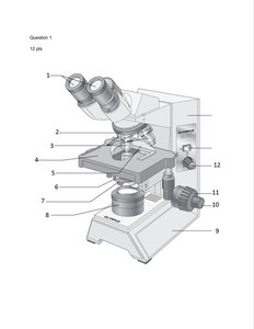
Try labeling and matching before revealing the answer!
Q6. True/False and correction: Microscope and slide handling procedures.
Background
Topic: Laboratory Safety & Technique
This question checks your understanding of proper microscope and slide handling, including cleaning, focusing, and using correct terminology.
Key Terms:
Special lens paper, scanning objective, coarse/fine adjustment, wet mounts, working distance, field of view.
Step-by-Step Guidance
Read each statement and determine if it is true or false based on lab protocols.
If false, identify the incorrect part and write the correct term or phrase (e.g., "use special grit-free lens paper" instead of "any soft tissue").
Review why each correction is necessary for proper microscope use and safety.
Practice these procedures in the lab to reinforce your understanding.
Try correcting the statements before revealing the answer!
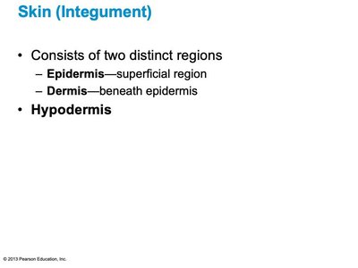
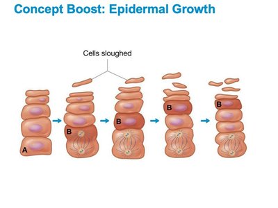
Q7. Integumentary System: Structure, layers, and cell types of the skin.
Background
Topic: Integumentary System Anatomy
This question covers the structure and function of the skin, including the epidermis, dermis, and hypodermis, as well as the types of cells found in each layer.
Key Terms:
Epidermis: Outermost layer, stratified squamous epithelium.
Dermis: Deeper layer, connective tissue, contains blood vessels and nerves.
Stratum basale, stratum corneum, melanocytes, keratinocytes.
Step-by-Step Guidance
Identify the main layers of the skin and their order from superficial to deep.
Recall the types of cells found in each layer and their functions (e.g., melanocytes produce melanin).
Understand the difference between the epidermis and dermis in terms of structure and function.
Practice labeling diagrams of the skin and describing the role of each cell type.
Try labeling and describing before revealing the answer!
Q8. Identify and describe the functions of skin glands and accessory structures.
Background
Topic: Skin Glands & Accessory Structures
This question focuses on the types of glands (sweat and sebaceous) and accessory structures (hair, nails) found in the skin, as well as their functions.
Key Terms:
Sudoriferous (sweat) glands: Eccrine and apocrine types.
Sebaceous (oil) glands: Produce sebum.
Hair, nails: Structure and function as skin appendages.
Step-by-Step Guidance
List the types of glands found in the skin and describe their secretions and functions.
Identify the difference between eccrine and apocrine sweat glands.
Describe the structure and function of hair and nails as derivatives of the epidermis.
Practice identifying these structures on diagrams and explaining their roles in homeostasis.
Try describing the functions before revealing the answer!
Final Answers (Selected Examples)
Q1: Brachial = arm, Cervical = neck, Digital = fingers/toes, etc.
Q4: Negative feedback reverses the original stimulus (e.g., body temperature regulation), while positive feedback amplifies it (e.g., blood clotting).
Q7: The main layers of the skin are the epidermis (outer), dermis (middle), and hypodermis (deepest, not technically part of the skin). The stratum basale is the deepest layer of the epidermis, containing melanocytes that produce melanin to protect against UV radiation.
Q8: Eccrine glands are the most widespread sweat glands and function in thermoregulation. Sebaceous glands produce sebum to lubricate and waterproof the skin and hair.
